fetching data ... 
Background: Golimumab is a human anti-TNF monoclonal antibody that has shown efficacy in RA. In the RA pathophysiology, the elevation of TNF modulates the cellular immune response, which leads to a sustained activation of T-cell response; however, it is unknown whether anti-TNF interferes with the immunophenotype of T-cell.
Objectives: To evaluate activation (CD25, CD69) and exhausted (PD-1, TIM-3, LAG-3, CTLA-4) markers of T-cells in RA patients treated with Golimumab.
Methods: We included 14 patients with RA diagnosis (Criteria ACR/EULAR 2010), with moderate to severe activity; 11 non-responders to synthetic DMARDs (NR-DMARDs) and 3 responders to synthetic DMARDs with pharmacological toxicity (R-DMARDs). All patients were treated with Golimumab during 24 weeks. The questionnaire SF-36, DAS28-CRP, power Doppler signal and expression of CD25, CD69, PD-1, TIM-3, LAG-3 and CTLA-4, in T-cell were evaluated at 0 and 24 weeks.
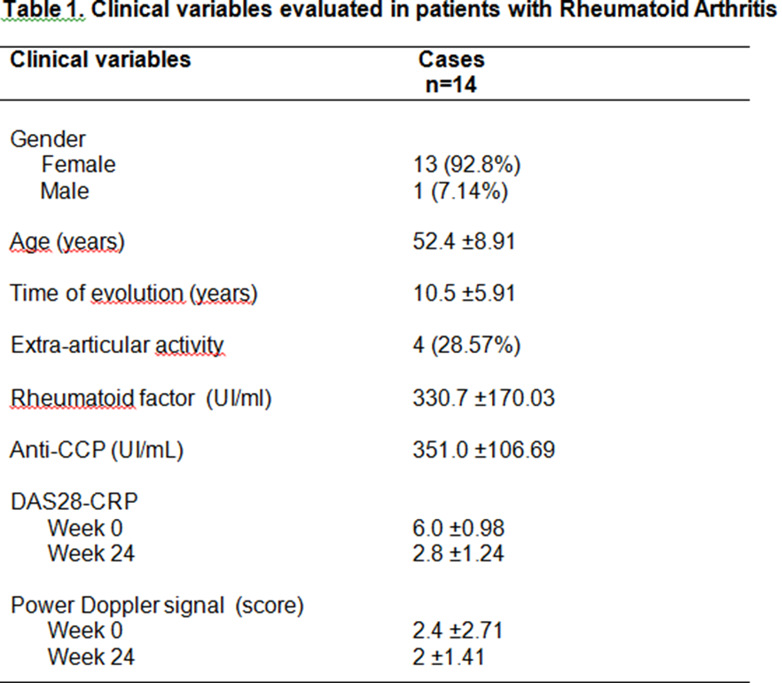
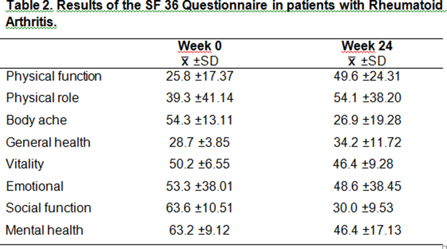
Results: Clinical variables evaluated in patients with Rheumatoid Arthritis are shown in tables 1 and 2.
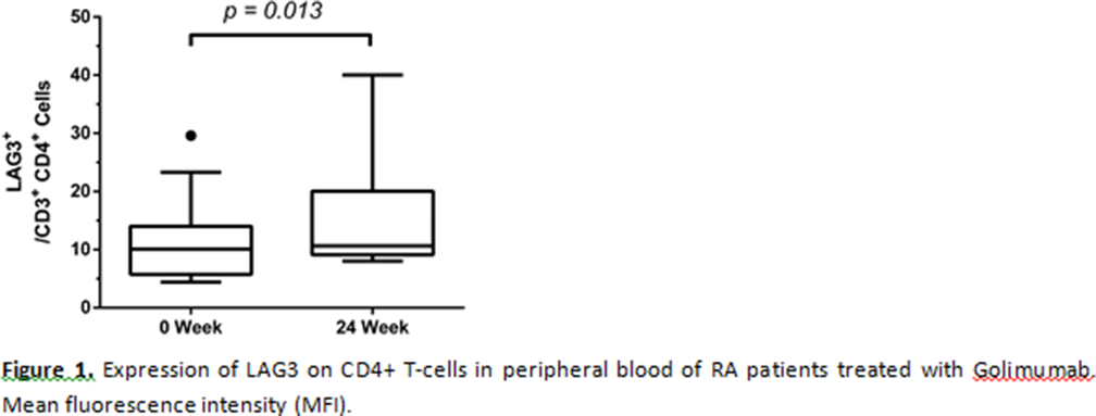
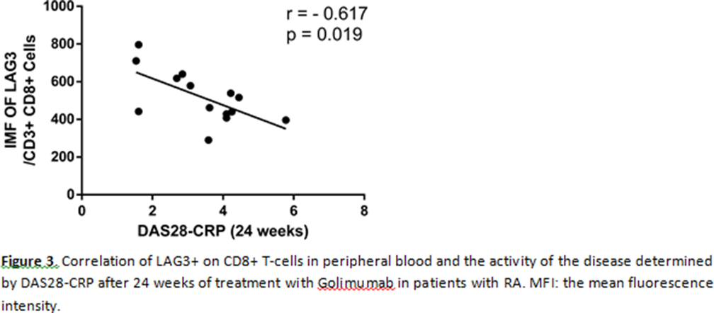
The frequency of LAG-3+ on CD4+ T-cells increased after 24 weeks of treatment with Golimumab (p = 0.013) (
Conclusion: Golimumab treatment increased the expression of LAG-3 in T-cells, suggesting a negative regulator of antigen presentation of T-cells.
REFERENCES:
[1] Tahir Z, Kavanaugh A. The role of golimumab in inflammatory arthritis. A review of the evidence. Ther Adv Musculoskel Dis2018;9:181–194.
[2] Cuda C, Pope R, Perlman H. The inflammatory role of phagocyte apoptotic pathways in rheumatic diseases. Nat rev Rheumatol2016;12: 543-555.
[3] Wherry E, Kurachi M. Molecular and celular insights into T cell exhaustion. Nat rev Inmunol 2015;15: 486-496.
[4] Burmester G, Feist E, Dörner T. Emerging cell and cytokine targets in rheumatoid arthritis. Nat rev Inmunol2013;15:1-10.
[5] McKinney E, Lee J, Jayne D, Lyons P. T-cell exhaustion, co-stimulation and clinical outcome in autoimmunity and infection. Nat rev Inmunol2015;523: 612-616.
Disclosure of Interests: None declared
DOI: 10.1136/annrheumdis-2019-eular.7666