fetching data ... 
Background: Peripheral neuropathy is one of the most frequent extraglandular manifestations of primary Sjögren’s syndrome (pSS). The diagnosis of peripheral neuropathy complications of pSS is based primarily on careful neurologic examination and electrodiagnostic tests. The value of ultrasound in peripheral nerve has been recognized. However, little clinical researches have focused specifically on cutaneous nerve of pSS.
Objectives: To evaluate the morphological changes of sural nerve in patients with pSS by high-frequency ultrasound.
Methods: The prospective study subjects consisted of 31 consecutive pSS patients underwent sural nerve biopsy and 30 healthy volunteers as controls. The ultrasonic presentations of the fascicle, perineurium, epineurium of sural nerve were observed, and the cross-sectional areas (CSA) of the sural nerves was measured.
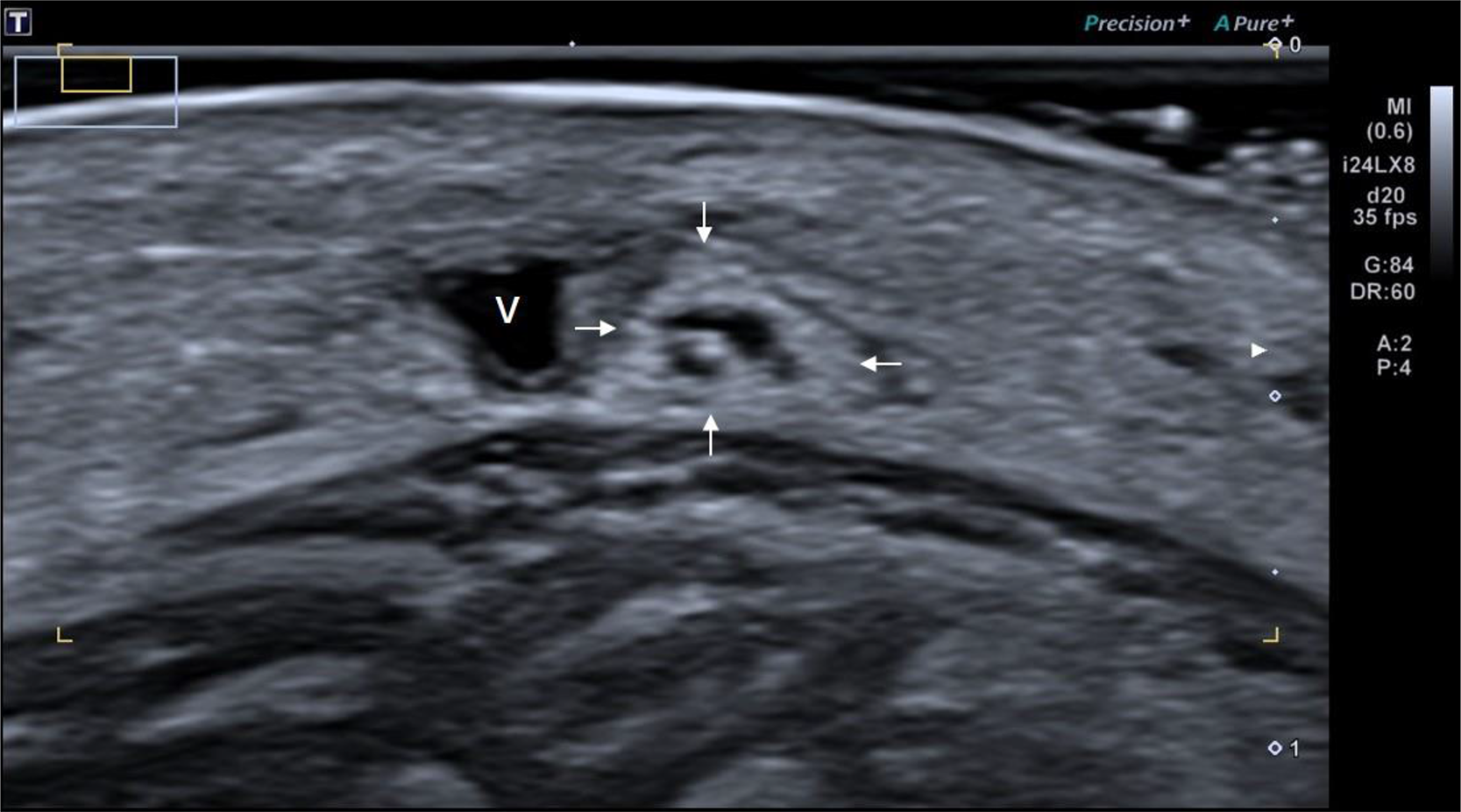
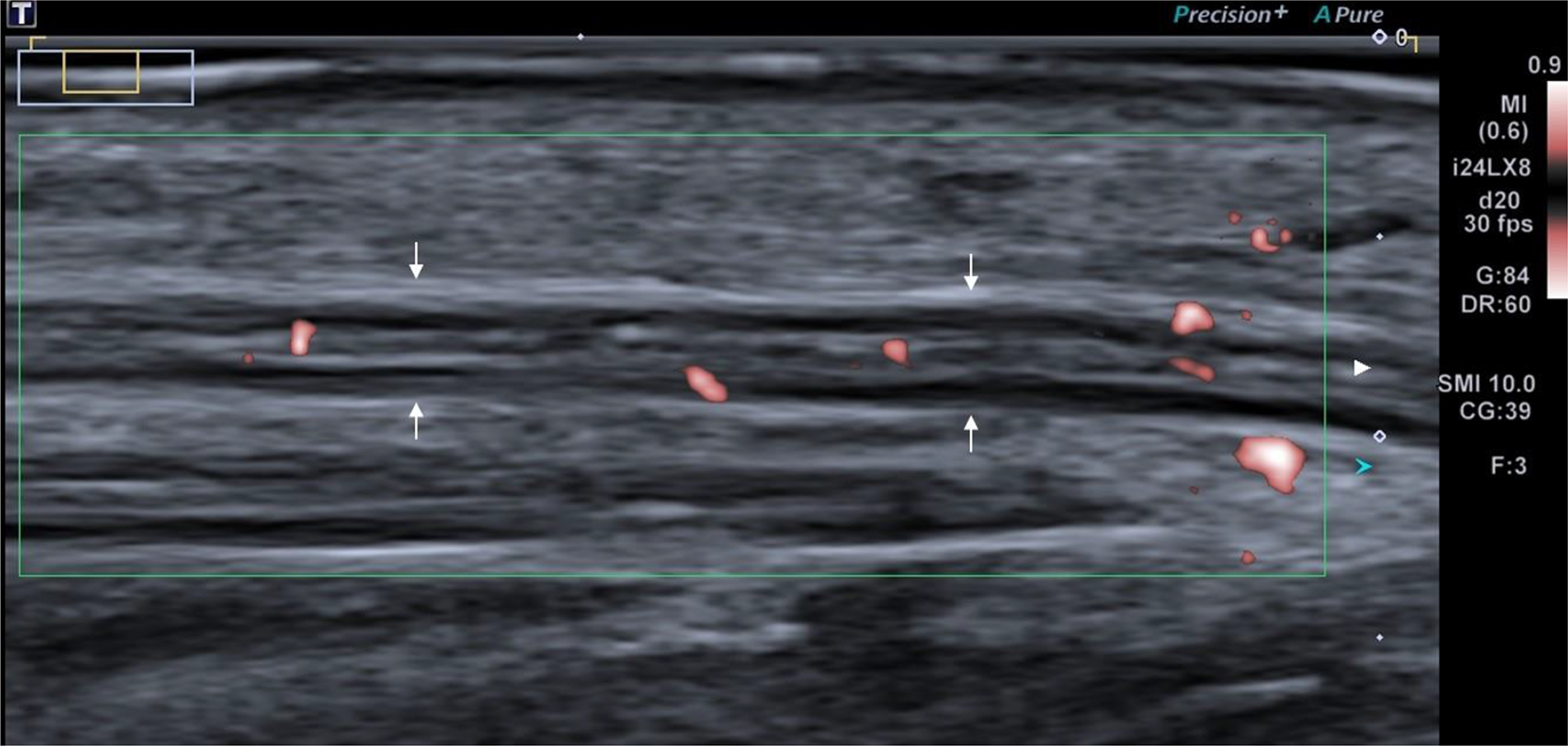
Results: Among the 21 sural nerves confirmed by pathology, all showed the thickening of the perineurium and epineurium (Figure 1-2), and abnormal blood flow signal in perineurium or epineurium in 14 cases (Figure 2). The mean CSAs were (1.41±0.44) mm2 for the control group, and (1.58±0.48) mm2 for the case group (P>0.05). In addition, the abnormal blood flow signal in sural nerve correlated with disease activity.
Conclusion: This study indicated that high-frequency ultrasound may be a valuable tool for evaluating cutaneous nerve neuropathy of Sjogren’s syndrome patients.
REFERENCES:
[1]Vitali C, Bombardieri S, Jonsson R, Moutsopoulos HM, Alexander EL, Carsons SE, et al. Classification criteria for Sjögren’s syndrome: a revised version of the European criteria proposed by the American-European Consensus Group. Ann Rheum Dis. 2002;61(6):554-8.
[2]Terrier B, Lacroix C, Guillevin L, Hatron PY, Dhote R, Maillot F, et al. Diagnostic and prognostic relevance of neuromuscular biopsy in primary Sjögren’s syndrome-related neuropathy. Arthritis Rheum.2007;57(8):1520-9.
[3]McCoy SS, Baer AN. Neurological Complications of Sjögren’s Syndrome: Diagnosis and Management. Curr Treatm Opt Rheumatol. 2017;3(4):275-88.
[4]Carvajal Alegria G, Guellec D, Devauchelle-Pensec V, Saraux A. Is there specific neurological disorders of primary Sjögren’s syndrome? Joint Bone Spine. 2015;82(2):86-9.
Transverse sonograms of the sural nerve (arrows) V: indicates lesser saphenous vein
Longitudinal sonograms of the sural nerve (arrows) The sonogram of sural nerve showed abnormal blood flow signal. V indicates lesser saphenous vein.
Acknowledgments: This work was partly supported by National Natural Science Foundation of China (No. 81701712).
Disclosure of Interests : None declared