
Background: Serum autoantibodies closely reflect patterns of organ involvement and disease progression in systemic sclerosis (SSc). The entire autoantibody profile is less well defined in many cohorts and the data regarding their clinical associations and frequencies is limited.
Objectives: To determine the autoantibody profile of patients with SSc, as well as their clinical associations, in well-characterized inception- cohort with disease duration less than 3 years.
Methods: Serum samples of 100 patients out of 105 enrolled in the study were analyzed for ANA patterns with indirect immunofluorescence (IIF) assay using HEp-20-10/primate liver mosaic IIFT kit. Sera of 96 patients were subjected to commercial line immunoassay to quantify autoantibodies against 13 different autoantigens.
Results: 92 (92%) out of 100 patients were positive for ANA by IIF (
Demographic, clinical and laboratory characteristics of the SSc patients.
| Sex | |
| Female N, % | 91 (86.7%) |
| Male N, % | 14 (13.3) |
| Female-to-male ratio | |
| Age, mean±SD years | 48.6±12.7 |
| Disease duration, mean±SD years | 2±1.4 |
| Disease classification N, % | |
| Diffuse | 39 (36.5%) |
| Limited | 65 (62.5%) |
| Sine scleroderma | 1 (1%) |
| Interstitial lung disease | 37 (34.3%) |
| Pulmonary arterial hypertension | 3 (2.8%) |
| Scleroderma renal crisis | 0 |
| Digital ulcer | 14 (13.3%) |
| Raynaud phenomenon | 105(100%) |
| Telengiectasia | 31 (28.8%) |
| Calcinosis | 1 (1%) |
| Malignancy | 3 (2.9%) |
| Antinuclear antibody profile N*, % | |
| Positive | 92 (92%) |
| Staining pattern | |
| Speckled | 65 (65%) |
| Nucleolar | 13 (13%) |
| Centromere | 29 (29%) |
| Homogeneous | 3 (3%) |
| Reticular | 3 (3%) |
Numbers and combinations of autoantibodies identified in the 96 SSc patients.
| Topo-I | CENP | RNAP III | Fibrillarin | NOR90 | Th/To | Pm/Scl | Ku | PDGFR | Ro52 | |
|---|---|---|---|---|---|---|---|---|---|---|
| Topo-I | 22 | 1 | 7 | 0 | 1 | 5 | 9 | 5 | 0 | 7 |
| CENP | 12 | 2 | 0 | 2 | 2 | 2 | 2 | 0 | 11 | |
| RNAPIII | 4 | 0 | 1 | 2 | 1 | 3 | 0 | 7 | ||
| Fibrillarin | 0 | 0 | 0 | 0 | 0 | 0 | 0 | |||
| NOR90 | 0 | 1 | 1 | 0 | 0 | 2 | ||||
| Th/To | 0 | 4 | 2 | 0 | 2 | |||||
| Pm/Scl | 4 | 3 | 0 | 6 | ||||||
| Ku | 1 | 0 | 2 | |||||||
| PDGFR | 0 | 0 | ||||||||
| Ro52 | 2 | |||||||||
| Single positive | 22 | 12 | 4 | 0 | 0 | 0 | 4 | 1 | 0 | 2 |
| Total | 45 | 27 | 18 | 0 | 5 | 8 | 17 | 9 | 0 | 24 |
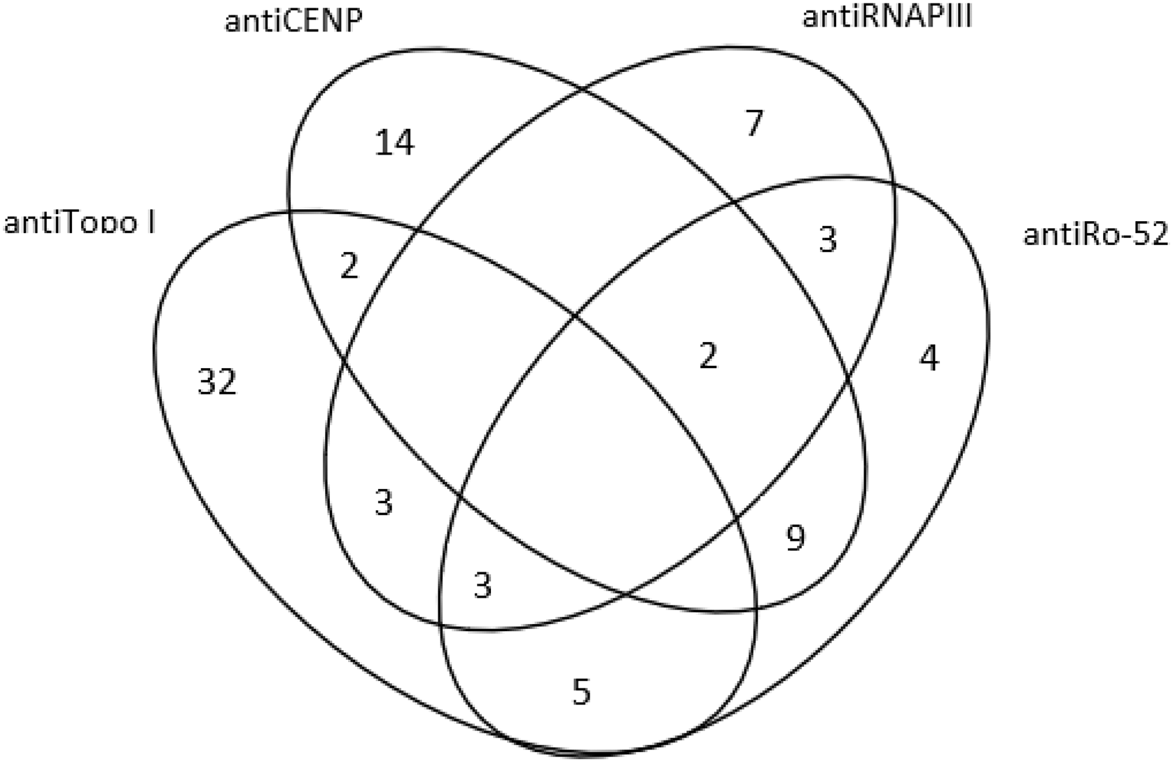
Diagram of disease-related antibodies against the four main autoantibodies [anti-centromere (antiCENP) anti-Topoisomerase I (antiTopo I), anti-RNA polymerase III (antiRNAP III) and anti-Ro52).

Conclusion: We presented the clinical and serologic features of the Turkish SSc patients from a new inception cohort. Clinical features of the SSc patients with single or multiple antibody positivity were not different.
REFERENCES:
None
Disclosure of Interests: None declared