fetching data ... 
Background: Hydatid cyst disease is a zoonosis caused by Echinococcus granulosus . The parasite implants most commonly in the liver and lung. Musculoskeletal involvement is rare.
Objectives: To describe clinical and imaging presentation of spinal and muscular hydatidosis.
Methods: We report two cases of chronic low back pain caused by hydatid cyst.
Results: Case 1: A 36-year-old woman living in a rural area presented with a three-years history of lumbar back pain complicated with right-sided sciatica and intermittent episodes of urinary retention. The pain was gradually worsening and resisting conventional analgesics and physiotherapy. On physical examination, she had tenderness of the lumbar back and left sacroiliac joint with limited mobility. Biological findings were normal and X-ray features showed osteolytic lesions of L5 left side. The magnetic resonance imaging (MRI) results revealed septated multilocular hydatic cysts with high signal intensity in T2-weighted images and low signal intensity in T1-weighted images along L5 and the sacrum left side. Serological test for hydatid disease was positive. The treatment was wide surgical resection combined with Albendazole (400 mg/day) for one year postoperatively.
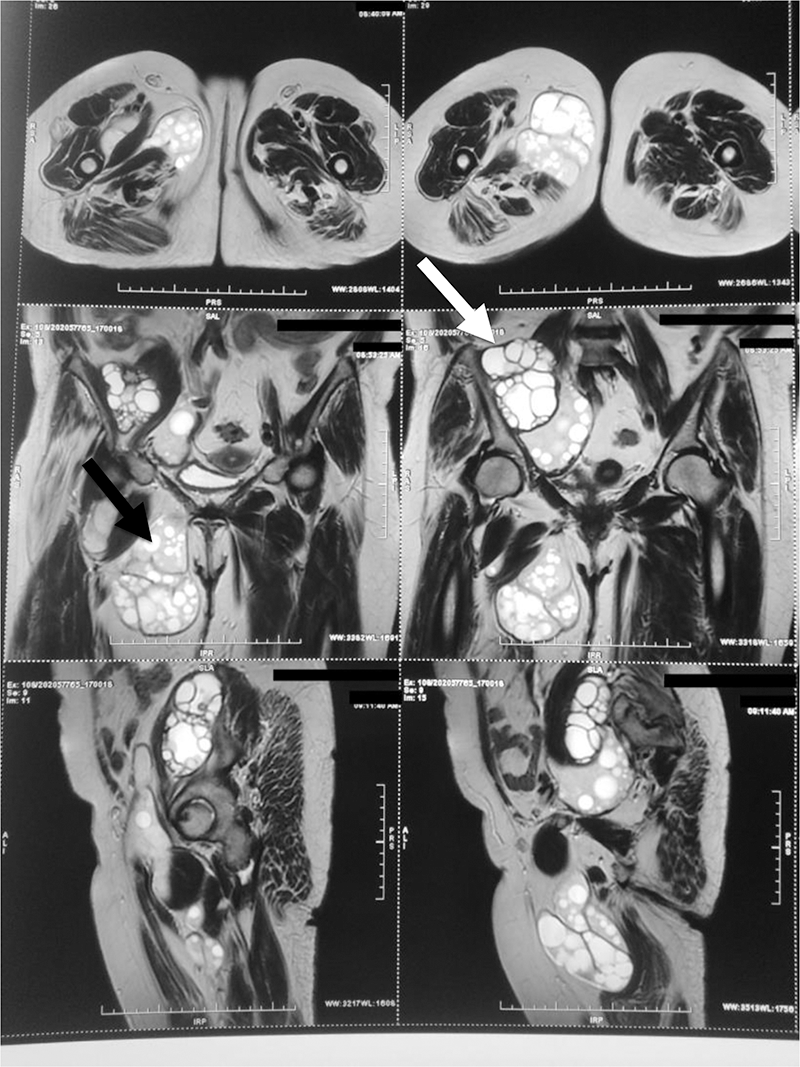
Case 2: A 62-year-old woman presented with a one-year history of lumbar back pain with gait instability and right flank swelling. Biological findings and X-ray features were normal. The patient first was explored by uro-CT to assess for nephrolithiasis and it revealed retro and sub-peritoneal two multilocular hydatic cysts (60x80mm, 36x35mm) along right iliopsoas and adductor muscles. MRI showed same cystes described on the CT (
MRI showing two multilocular cysts along right iliopsoas (white arrow) and adductor muscles (black arrow).
Conclusion: In endemic areas, bone and muscle involvement from echinococcosis should be considered during clinical investigation of chronic lumbar pain.
Disclosure of Interests: None declared