fetching data ... 
Background: Type I Interferons (IFN) are central players in the pathogenesis, disease activity and evolution of several autoimmune conditions. To date, a limited number of evidences is available on the specific role of IFN activation in antiphospholipid antibodies (aPL) positive patients, including aPL carriers, primary antiphospholipid syndrome (PAPS) and those APS subjects who presented with an associated autoimmune disease (secondary APS, SAPS), such as systemic lupus erythematosus (SLE).
Objectives: The aim of this study was to evaluate the differential expression of IFN stimulated genes (ISG) among different subsets of aPL positive subjects and SLE patients.
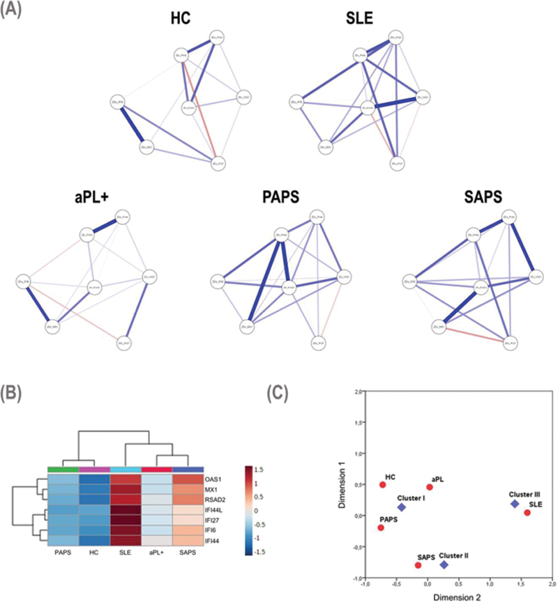
Methods: For the purpose of the study, a total of 112 patients attending the San Giovanni Bosco Hospital (Turin, Italy) were enrolled, including 31 PAPS, 25 SAPS, 27 SLE patients without aPL, 29 aPL carriers (mean age 48.3±13.3 years, 76% female) 1,2 . Nineteen subjects were also recruited as healthy controls (HCs). Complete demographic, clinical, and laboratory data were collected at the time of the inclusion. Gene expression was evaluated by RT-PCR in whole blood for the following genes: IFI6, IFI44, IFI44L, MX1,IFI27, OAS1 and RSAD2. Normalized gene expression levels (Z-scores) were averaged into a global IFN signature (IFN score). Differences were measured by Kruskal-Wallis tests and associations among genes were studied by cluster and correspondence analyses. Correlations among genes were plotted by network analyses.
Results: An overall activation of ISG was noted across APS subsets, but certain differences were noted among genes. Whereas some ISG were already upregulated in the aPL positive group compared to HC (IFI44, IFI44L, MX1, IFI27, OAS1 and RSAD2, all p<0.050), other ISG were only in increased SLE (IFI6), MX1 differed between SLE and SAPS, and IFI27 and OAS1 showed differences between PAPS and SAPS. The composite IFN score revealed quantitative differences in the IFN pathway activation across APS subsets, being elevated in aPL carriers/PAPS groups compared to HCs (both p<0.050) and increasing in SAPS (p<0.010) and SLE (p<0.001) groups. Network analyses (
Conclusion: An overall IFN pathway activation has been observed in aPL positive patients and across all APS subsets. Qualitative and quantitative differences across the APS spectrum can be identified, leading to the identification of distinct IFN signatures with different clinical value.
REFERENCES:
[1]Miyakis S, et al. J Thromb Haemost (2006). 2. Aringer M, et al. Arthritis Rheumatol (2019).
Disclosure of Interests: None declared