fetching data ... 
Background: Systemic lupus erythematosus (SLE) is an inflammatory autoimmune disease, involving the development of autoreactive cells and autoantibodies. Natural Killer (NK) cells are innate immune cells that mediate the interaction between the innate and adaptive immune system, however their role in SLE is incompletely understood. SLE NK cells are decreased in peripheral blood, exhibit reduced cytotoxicity, and impaired cytokine production (1, 2). Furthermore, SLE NK cells present phenotypic alterations: increased expression of CD38 and altered upregulation of SLAMF7 after activation (3). To date, few studies evaluated the molecular mechanisms underlying NK cell dysfunction in SLE.
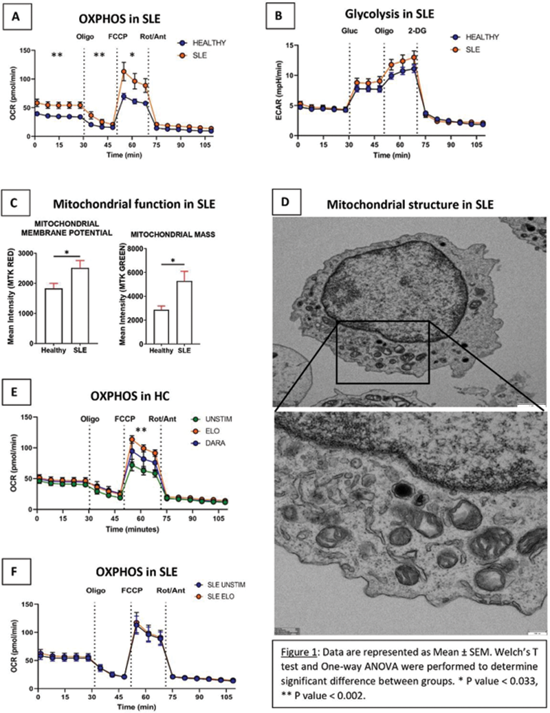
Objectives: We examined immunometabolic alterations of SLE NK cells. First, we characterized the cellular metabolism of SLE NK cells by assessing glycolysis and oxidative phosphorylation (OXPHOS) at basal level. Then, we evaluated how cellular metabolism can be manipulated to enhance NK cell function. In this perspective, we examined how the ligation of CD38 with daratumumab (DARA) and SLAMF7 with elotuzumab (ELO) modulate glycolysis and OXPHOS.
Methods: NK cells of cryopreserved PBMC from SLE patients were isolated. Glycolysis and OXPHOS were studied using XF e 96 Seahorse. Expression of metabolic receptors (CD71, GLUT-1, CD98), mitochondrial function (mitochondrial membrane potential, mass) and calcium influx were investigated by FACS. Mitochondrial structure was evaluated by electron and confocal microscopy.
Results: First, we examined the cellular metabolism of SLE NK cells compared to healthy cells. We observed that OXPHOS is significantly increased in SLE NK cells (
Second, we examined how ligation of DARA and ELO influences the metabolism of healthy NK cells. Our data showed that ELO primarily enhances NK cell OXPHOS (
Conclusion: Our data suggest that SLE NK cells exhibit alterations in cellular metabolism, primarily involving mitochondrial respiration. In contrast, glucose metabolism is similar to that of healthy NK cells. Additionally, ELO and DARA mediate the activation of healthy NK cells through the engagement of different metabolic pathways: OXPHOS and glycolysis, respectively. Therefore, priming SLE NK cells with ELO is unable to adequately engage their dysfunctional mitochondrial respiration. These findings provide important insights on the alteration present in SLE NK cells and contribute to a better understanding of the pathogenesis of the disease.
REFERENCES:
[1]Spada R, Rojas JM, Barber DF. Recent findings on the role of natural killer cells in the pathogenesis of systemic lupus erythematosus. J Leukoc Biol. 2015;98(4):479-87.
[2]Park Y-W, Kee S-J, Cho Y-N, Lee E-H, Lee H-Y, Kim E-M, et al. Impaired differentiation and cytotoxicity of natural killer cells in systemic lupus erythematosus. Arthritis & Rheumatism. 2009;60(6):1753-63.
[3]Humbel M, Bellanger F, Fluder N, Horisberger A, Suffiotti M, Fenwick C, et al. Restoration of NK Cell Cytotoxic Function With Elotuzumab and Daratumumab Promotes Elimination of Circulating Plasma Cells in Patients With SLE. Front Immunol. 2021;12:645478.
Acknowledgements: This study received funding from the Swiss National Science Foundation (Ambizione PZ00P3_173950 to DC).
Disclosure of Interests: None declared