fetching data ... 
Background: Neuronal damage in systemic lupus erythematosus (SLE) is common, but the extent and mechanisms are unclear [1-2]. Neurofilament light (NfL) concentrations rise in plasma and cerebrospinal fluid (CSF) during neuronal damage and reach abnormal levels in various neurological disorders [3]. NfL is sparsely studied in SLE [4-7].
Objectives: To explore plasma and CSF concentrations of NfL in SLE patients and investigate the associations between NfL and nervous system involvement, including cognitive dysfunction, imaging findings on magnetic resonance imaging (MRI), laboratory and clinical abnormalities, and to compare the NfL levels of SLE patients with those present in healthy controls.
Methods: In this cross-sectional study, 72 consecutive SLE out-patients and 26 healthy controls, all female, aged <55 years, underwent MRI and neurocognitive testing. NfL concentrations in plasma from all individuals and in CSF from 32 patients were measured with single-molecule array technology. Patients were assessed by a rheumatologist and neurologist to define neuropsychiatric involvement (NPSLE) according to three attribution models.
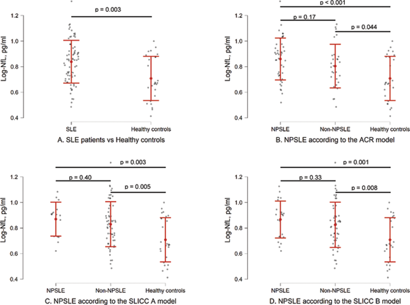
Results: Plasma and CSF NfL concentrations correlated strongly (r=0.72, p<0.001). Plasma NfL concentrations were higher in SLE patients, both with and without neuropsychiatric involvement, compared with healthy controls (
Plasma log-NfL concentrations between groups. The intervals illustrate means and standard deviations.
Conclusion: Higher plasma NfL concentrations in NPSLE and non-NPSLE patients may indicate a higher degree of neuronal damage in SLE in general, particularly in the lower age group, corresponding with cognitive impairment and organ damage development. NfL may serve as an indicator of neuronal damage in SLE in further studies.
REFERENCES:
[1]Jeltsch-David H, Muller S (2014) Neuropsychiatric systemic lupus erythematosus: Pathogenesis and biomarkers. Nature Reviews Neurology 10:579–596
[2]Hanly JG, Walsh NMG, Sangalang V (1992) Brain pathology in systemic lupus erythematosus. Journal of Rheumatology 19:732–741
[3]Gaetani L, Blennow K, Calabresi P, et al (2019) Neurofilament light chain as a biomarker in neurological disorders. Journal of Neurology, Neurosurgery and Psychiatry 90
[4]Trysberg E, Nylen K, Rosengren LE, Tarkowski A (2003) Neuronal and Astrocytic Damage in Systemic Lupus Erythematosus Patients with Central Nervous System Involvement. Arthritis and Rheumatism 48:2881–2887.
[5]Tjensvoll AB, Lauvsnes MB, Zetterberg H, et al (2020) Neurofilament light is a biomarker of brain involvement in lupus and primary Sjögren’s syndrome. Journal of Neurology.
[6]Lauvsnes MB, Zetterberg H, Blennow K, et al (2021) Neurofilament light in plasma is a potential biomarker of central nervous system involvement in systemic lupus erythematosus. Journal of Neurology 1:1–11.
[7]Engel S, Boedecker S, Marczynski P, et al (2021) Association of serum neurofilament light chain levels and neuropsychiatric manifestations in systemic lupus erythematosus. Therapeutic Advances in Neurological Disorders 14:175628642110514.
Disclosure of Interests: None declared