fetching data ... 
Background: Gouty arthritis (GA) is an autoinflammatory disease caused by the deposition of monosodium urate crystal (MSU) in the joints and surrounding tissues, which lead to a series of complex inflammatory cascade amplification reactions.The clinical symptoms of acute GA attack rapidly, but often alleviate spontaneously within 7 ~ 10 days, which is one of the significant characteristics different from other joint diseases or autoimmune diseases. However, the exact molecular mechanism of its inflammatory self limitation is still unclear. The phenotypic imbalance of Th1 / Th2 cells and the M1/ M2 polarization of macrophages may be involved in the inflammatory self limitation of gout [1] .Decoy receptor 3 (DCR3) can differentiate T cells into Th2 phenotype, promote M2 polarization of macrophages, and play the functions of immune regulation and repair [2] .DCR3 and its Signal Pathway are involved in the pathogenesis of tumors and a variety of autoimmune diseases, and have become an important research target of tumors and immune related diseases.However, studies on DcR3 related molecular pathway and GA are scarce, and the specific regulatory mechanism is unknown.
Objectives: To assess the contribution of DcR3 and its signal pathway to gout and the clinical importance of these genes in primary gouty arthritis.
Methods: The mRNA expression levels of DCR3 and its signal pathway(DR3, TL1A, Fas, FasL, Ligth, LigthR, LTgthRNA expression levels of DCR3 and its signal pathway(DR3gout and the clinical importance of these genes in primary gouty arthritis.nt research target of tumors and immune related diseases.However, studies on DcR3 related moathway expression levels and laboratory features was analyzed in GA patients.
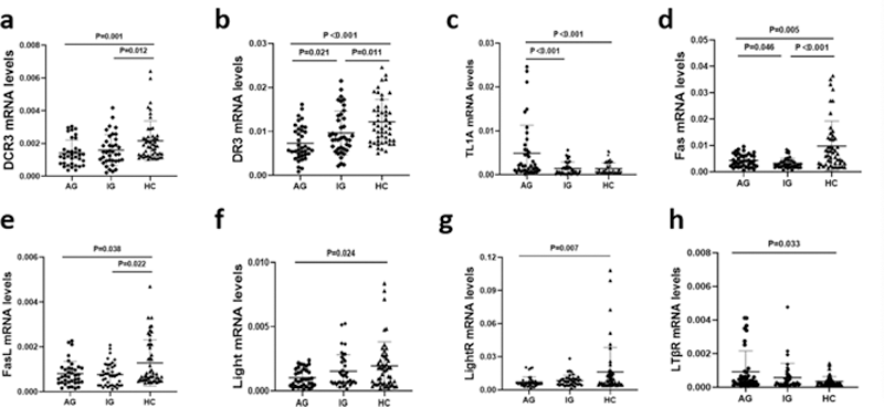
Results: The expression levels of DCR3, FasL were much lower in the AG and IG group than in the HC groups (p<0.05), and no significant difference was detected between AG and IG groups(P>0.05)(a,e). The expression levels of DR3 were much lower in the AG and IG group than in the HC groups (p<0.05), and much lower in the AG group than in the IG groups (p<0.05)(b). The expression levels of TL1A were much higher in the AG group than in the IG and HC groups (p<0.05), and no significant difference was detected between IG and HC groups(P>0.05)(c).The expression levels of Light, LightR were much lower in the AG group than in the HC groups (p<0.05), and no significant difference was detected between AG and IG groups, IG and HC groups(P>0.05)(f,g).The expression levels of LTlower in the AG and IG group than in the HC groups (p<0.05(p<0.05), and no significant difference was detected between AG and IG groups, IG and HC groups(P>0.05)(h).In GA patients, the levels of DcR3 related molecular pathway gene correlated with laboratory inflammatory and metabolic indexes.
Conclusion: Altered DCR3 and its signal pathway expression suggests that DCR3 related molecular pathway is involved in the pathogenesis of GA and participates in regulating inflammation and metabolism.
REFERENCES:
[1]Desai J, Steiger S, Anders HJ. Molecular Pathophysiology of Gout[J]. Trends Mol Med. 2017 Aug;23(8):756-768. DOI:10.1016/j.molmed.2017.06.005.
[2]Pan YG, Huang MT, Sekar P, et al. Decoy Receptor 3 Inhibits Monosodium Urate-Induced NLRP3 Inflammasome Activation via Reduction of Reactive Oxygen Species Production and Lysosomal Rupture[J]. Front Immunol. 2021 Mar 3;12:638676.DOI:10.3389/fimmu.2021.638676.
Relative Expression of DcR3 related molecular pathway gene in the PBMCs of Patients.The expression levels of DCR3, FasL were much lower in the AG and IG group than in the HC groups (p<0.05)(a,e). The expression levels of DR3 were much lower in the AG and IG group than in the HC groups (p<0.05), and much lower in the AG group than in the IG groups (p<0.05)(b). The expression levels of TL1A were much higher in the AG group than in the IG and HC groups (p<0.05)(c).The expression levels of Light, LightR were much lower in the AG group than in the HC groups (p<0.05)(f,g).The expression levels of LTβR were much higher in the AG group than in the HC groups (p<0.05)(h).
Acknowledgements: Institute of Research Center of Gout and Hyperuricemia of the Affiliated Hospital, North Sichuan Medical College
Disclosure of Interests: None declared