fetching data ... 
Background: Although the synovial subintima is one of the key points in joint pathology, its normal structure remains not fully understood.
Objectives: The morphology of the synovial subintima from birth till the 90th day was studied using the hip joints of intact Wistar rats.
Methods: Joints were harvested (on the first, seventh, 14th, 30th, 60th, and 90th day; six rats in each group, 36 in total), fixed, decalcified, and dehydrated. Paraffin-embedded tissue sections were stained with Malloryʼs trichrome, Hartʼs elastin, and Laidlaw staining. Light microscopy, morphometrical (intersection counting), and statistical methods with subsequent extrapolation to humans (Sengupta, 2013) were used. The obtained data were presented as percentages. The count of cells and extracellular matrix (ECM) components was performed in the larger part of the joint capsule which directly enclosed the articular cavity. Since the articular cavity and joint capsule in the anterior (ventral) part of the hip joint were not formed till the seventh day, they were not considered. The count in the femoral neck was not carried out due to some local features of the synovium (see below).
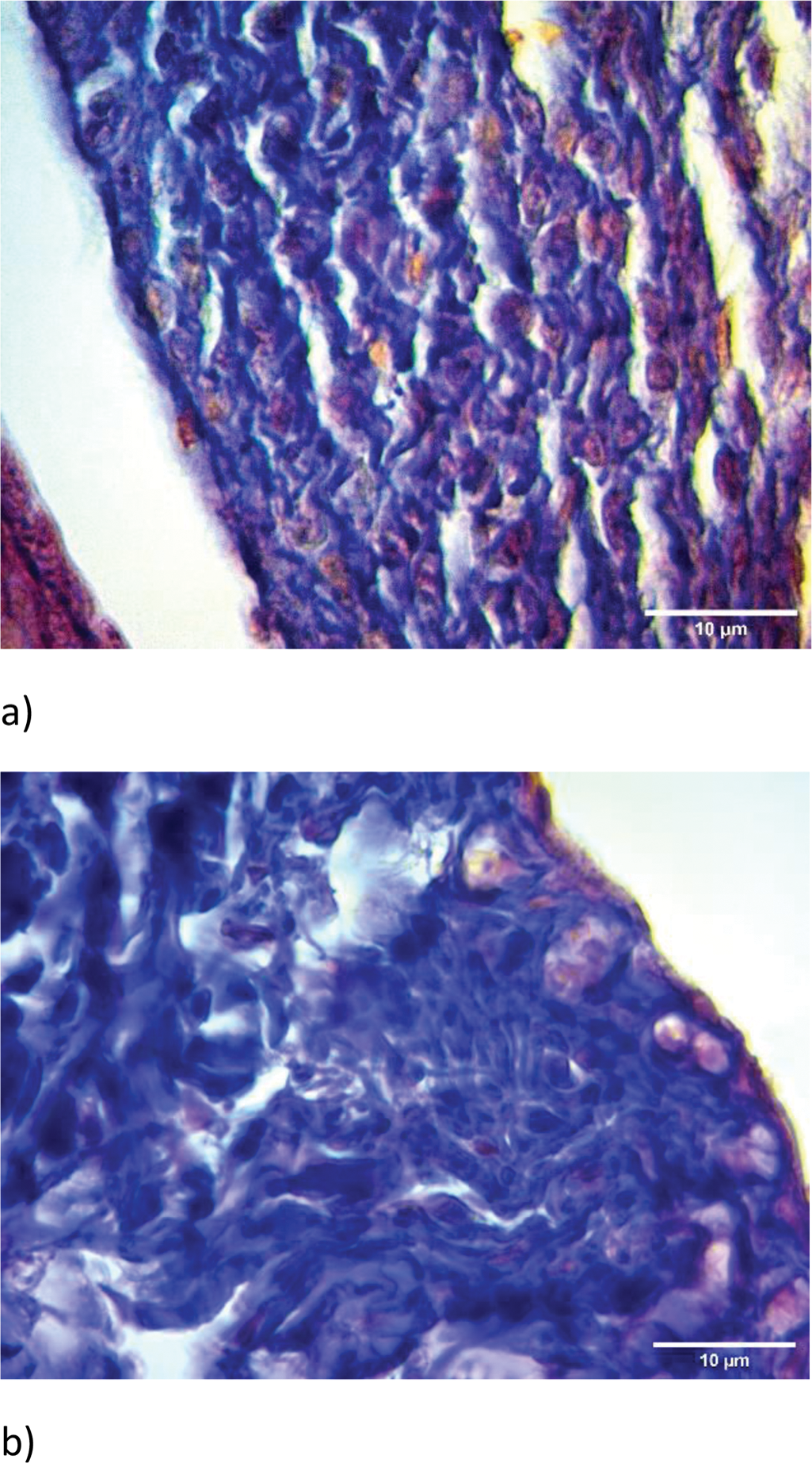
Results: The hip joint capsule presented the areolar synovium during the early postnatal period. With the development of fibrous components of the synovial subintima, its structure corresponded to the fibrous-areolar synovium. The overall quantity of collagen fibers increased gradually, exceeding on the 90th day (approximately corresponds to 18-19 human years) that of newborns more than twice (41.5±4.79 vs 80.5±7.79, p=0.0021). The number of arranged collagen fibers increased till the 30th day (10.65±1.25 vs 41.92±3.56, p<0.001; approximately corresponds to the 9th human year) and did not change significantly after that. The area of distribution of disarranged collagen fibers decreased till the 14th day (28.83±3.4 vs 18.5±1.67, p<0.001; approximately corresponds to the early infancy) with a subsequent increase, exceeding those of newborns on the 90th day almost 1.5 times (28.83±3.4 vs 39.7±3.1, p<0.001). In newborns, type III collagen prevailed over type I collagen. After the 14th day, type I collagen began to dominate. The number of elastic fibers increased till the 60th day (1.67±0.27 vs 45.6±4.22, p<0.001; approximately corresponds to the 16th human year). The amount of ground substance dramatically decreased by the seventh day (approximately corresponds to the early neonatal period of human life) about three times (29.6±3.14 vs 11.75±1.37, p<0.001) and did not change substantially after that. This is probably due to an increasing distribution density of fibers, reflecting an increase in the motor activity of rats. The area occupied with vessels increased till the 30th day (3.15±0.41 vs 6.9±0.39, p<0.001) with a subsequent gradual decrease till the 90th day (6.9±0.39 vs 4.75±0.43, p<0.001). The maximum number of cells was on the first and seventh days (26.58±1.93 and 26.92±1.36). Subsequently, it gradually decreased. The number of cells on the 90th day was almost four times less compared to newborns (26.58±1.93 vs 6.65±0.88, p<0.001). This partly confirms the statement that synovium is a relatively acellular structure (Smith 2011).
From the 30th day, an abundance of vascular bed and fatty tissue in the superficial layers of the synovial subintima which covers the femoral neck was observed. From the 60th day, it formed articular fat pads that would play an important role as a thermal insulator and shock absorber. In the deeper layers of the femoral synovial subintima, there was a predominance of arranged bundles of argyrophilic fibers. They formed periosteal Sharpey’s fibers of the femoral periosteum. The cranially directed fibers attached to the articular cartilage of the femoral head.
Conclusion: The morphogenesis of the hip joint capsule in the postnatal period reflects complex cell-cell and cell-ECM interactions and is accompanied by a decrease in the number of cells and ground substance, an increase in the number of fibers with an undulatory pattern in the distribution of vessels in it.
Normal synovium on the first (a) and 90th day (b), Mallory’s trichrome stain, x100.
REFERENCES: NIL.
Acknowledgements: NIL.
Disclosure of Interests: None declared.