fetching data ... 
Background: Glycoconjugates, in particular galactose-specific, contribute significantly to fundamental biological functions, such as cell differentiation, cell-cell and cell-matrix interaction which are of great importance for the normal functioning of the joint. Moreover, lectins play a key role in innate immunity by recognizing a variety of pathogens. The abnormal distribution of those is associated with the development of different joint inflammatory diseases. Their distribution in the normal articular cartilage remains insufficiently studied.
Objectives: Distribution of galactose-specific lectins was studied using the hip joint of intact Wistar rats (on the first, seventh, 14th, 30th, 60th, and 90th day; six rats in each group, 36 in total).
Methods: Paraffin-embedded tissue sections were stained with Helix pomatia agglutinin (HPA), peanut agglutinin (PNA), and soybean agglutinin (SBA) conjugated with horseradish peroxidase (HRP). The intensity of staining was evaluated semiquantitatively (from 0 to ++++).
Results: The most superficial zone (MSZ) revealed strong expression in the outermost and innermost sublayers; their distribution did not change significantly throughout the observation period. (Fedotchenko, 2023).
From birth to the seventh day, the tangential zone showed almost no HPA- and PNA-binding sites. The SBA revealed no affinity on the first day and a weak staining (+) on the seventh day. On the 14th day, there was no staining for the HPA, a weak (+) staining for the SBA, and a strong expression (+++) of receptors for the PNA.
From birth till the 14th day, the middle zone showed a weak (+) expression of studied receptors; the basal zone stained weakly (+) with the PNA and moderately (++) with the HPA and SBA.
The marginal cartilage showed no lectin-binding sites till the seventh day. On the 14th day, it showed a strong (+++) expression for the PNA and SBA and no affinity to the HPA.
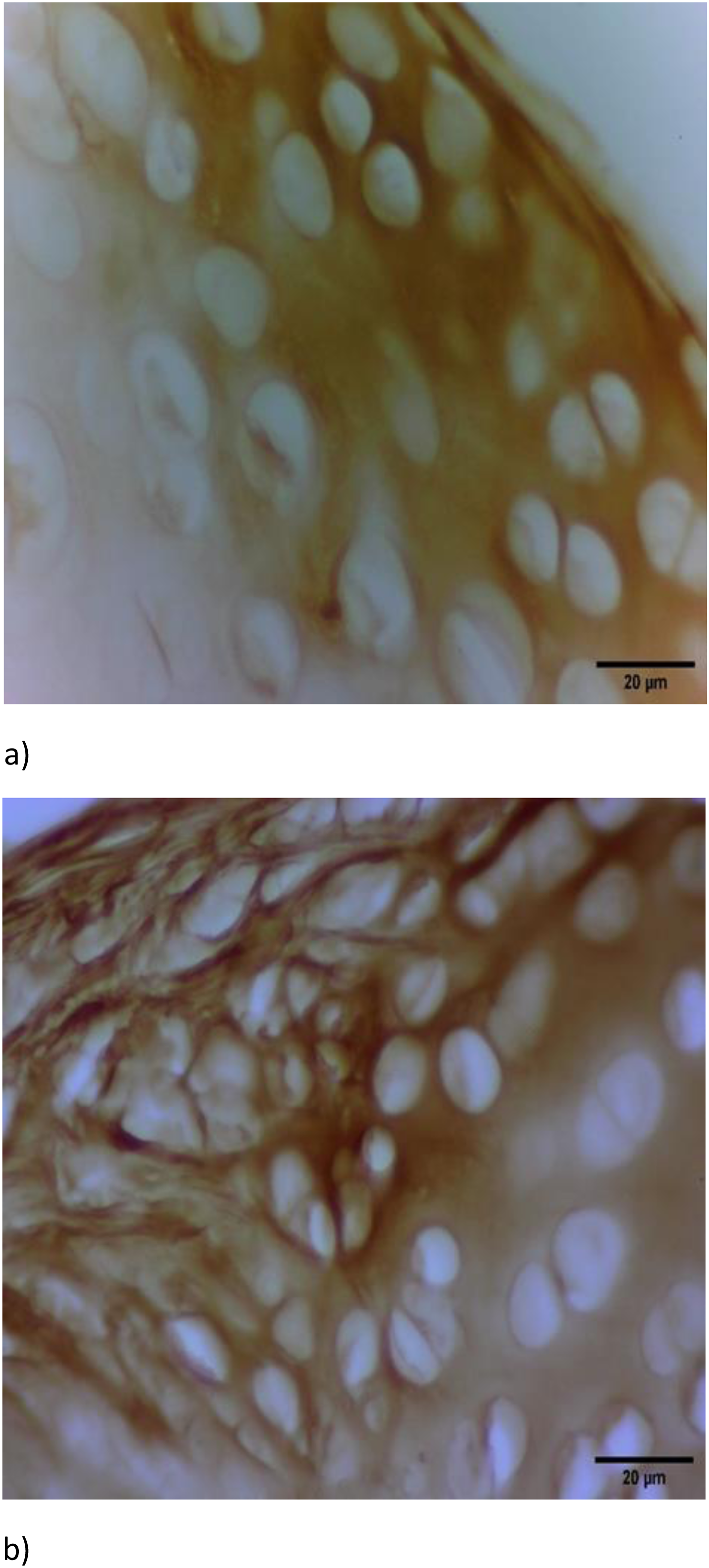
From the 30th day, the tangential zone showed almost no affinity to the HPA. At the same time, a robust expression (++++) of receptors for the PNA and SBA on the 30th day (Figure 1a) and a weak reaction (+) from the 60th day could be seen. Most of the middle zone showed almost no lectin affinity to the HPA and weak staining (+) for the PNA and SBA. And still, at the same time, single foci of irregular polygonal shape with strong lectin affinity (+++) to the studied lectins could be traced here. The deep zone and growth plate (proliferative, hypertrophic zones, and the calcified cartilaginous spicule) showed from the 30th day a strong expression (from +++ to ++++) to all studied lectins. The resting zone did not significantly differ in the intensity of staining from the middle zone of the articular cartilage. From the 30th day, the marginal cartilage showed no reaction to the HPA, but a strong affinity (from +++ to ++++) for the PNA and SBA (Figure 1b). The nuclei of chondrocytes showed various affinities (from weak (+) to strong (+++)). The well-defined cartilage lacuna was strongly stained (from +++ to ++++). In the distribution of lectins between the femoral head and the acetabulum, no substantial differences were observed.
Conclusion: With the development of the joint, the most lectin-binding affinity could be traced in the tangential zone, growth plate, cartilage lacunae, cell nucleus, and marginal cartilage, the less – in the middle zone. The highest intensity of staining showed peanut (PNA) and soybean (SBA) agglutinins; the lowest – Helix pomatia agglutinin (HPA). The strong expression of lectins in the growth plate might reflect the intensity of mitotic and biosynthetic processes that occur in it. The pronounced expression of lectins in the „border zones“, especially in the marginal cartilage and MSZ, appears to play the role of an innate immunobiological barrier that binds and neutralizes genetically alien material (antigens). Knowledge of lectin distribution in the norm is of extreme importance in better understanding of joint pathology.
The femoral head on the 30th day, staining with SBA-HRP, x40; a) the most superficial, tangential, and middle zones of the articular cartilage; b) the marginal transitional zone.
REFERENCES: NIL.
Acknowledgements: NIL.
Disclosure of Interests: None declared.