fetching data ... 
Background: Although many studies have been conducted on osteoarhritis (OA), there is no curative treatment yet. In addition to the negative effects of the progressive OA process on the patient’s functional characteristics and daily living activities, it also places a serious burden on the global health system. Therefore, slowing down its progression is also an important issue. Many studies have been conducted on the development of OA due to vitamin K deficiency [1]. However, it is not yet clear which type of vitamin K has the effect on OA. A study conducted in 2013 found that patients who developed knee OA had lower vitamin K2 levels in the medial part of the femorotibial joint than in the lateral [2]. Based on these data, our study aimed to examine the effect of vitamin K2 supplementation on the progression of knee OA.
Objectives: It was aimed to examine the effect of oral vitamin K2 supplementation on OA progression in the OA model induced by monosodium iodoacetate in rats.
Methods: 24 male Sprague Dawley rats were included in the study. The rats were divided into 3 equal groups: sham group, control (OA) group, and treatment group. Sterile saline was applied intraarticularly to the right knee of the sham group and Monosodium Iodoacetate (MIA) molecule was applied intraarticularly to the right knee of the control and treatment groups to create an OA model. In addition to the standard diet, 8 micrograms (µg)/ day of vitamin K2 was given orally to the rats in the treatment group. After 28 days of follow-up, the rats were sacrificed. Right knee articular cartilage was examined histologically with Hematoxylin - Eosin and Safranin O and immunohistochemically with type II collagen alpha 1 and Matrix Metalloproteinase - 13 (MMP - 13). Mankin was used as a histologic scoring system and H - Score scoring system was used for the evaluation of immunohistochemical data.
Results: Histologic examinations showed that the cartilage was more regular and the cartilage thickness was higher in the treatment group than in the control group and there were no synovial tissue remnants (Figure 1).
Images of groups staining with Safranin-O (a. sham group, b. control (OA) group, c. treatment group)
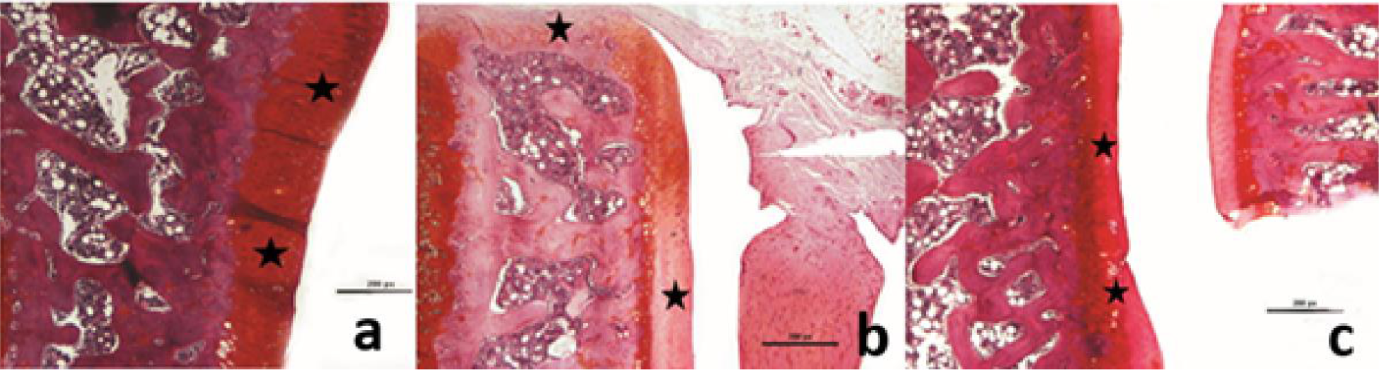
Mankin score data showed significantly lower results in the treatment group (4.25 ± 0.83) compared to the control group (11.10 ± 0.83). Immunohistochemical examinations revealed more intense type II collagen staining and less MMP-13 staining in the treatment group compared to the control group. According to H-Score data, type II collagen level was significantly higher in the treatment group (35.00 ± 7.60) compared to the control group (6.30 ± 2.31), while MMP-13 level was significantly lower in the treatment group (28.80 ± 8.30) compared to the control group (71.30 ± 2.31).
Conclusion: It was found that OA progression could be slowed down by oral vitamin K2 administration in the OA model induced by MIA in rats. This is the first study in which vitamin K2 was found to be effective on OA progression.
REFERENCES: [1] Chin K. Y. (2020). The Relationship between Vitamin K and osteoarthritis: A Review of Current Evidence. Nutrients, 12(5), 1208.
[2] Ishii, Y., Noguchi, H., Takeda, M., Sato, J., Yamamoto, N., Wakabayashi, H., Kanda, J., & Toyabe, S. (2013). Distribution of vitamin K2 in subchondral bone in osteoarthritic knee joints. Knee surgery, sports traumatology, arthroscopy: official journal of the ESSKA, 21(8), 1813–1818.
Acknowledgements: NIL.
Disclosure of Interests: None declared.