
Background: There is no standardization of minor salivary gland biopsy (MSGB) in patients with Sjögren’s syndrome (SS) for accurate interpretation of immunohistochemistry (IHC) related findings. IHC could characterize the phenotype of lymphocytes present in an MSGB and be useful in defining prognosis.
Objectives: The aim was to describe the histopathologic and IHC variables in patients with sicca syndrome and MSGB with focus score (FS) ≥1.
Methods: Retrospective observational study of patients with sicca syndrome. Clinical, histopathological and IHC characteristics of MSGB were described using red (CD8 T cells) and brown (CD4 T cells) chromogen-based staining. CD20:CD3 and CD4:CD8 markers were obtained using Easy Scan Pro 6 -MOTIC® and QuPath® software. Descriptive statistics: qualitative variables (Chi2 or Fischer’s exact test) and quantitative variables (according to normality
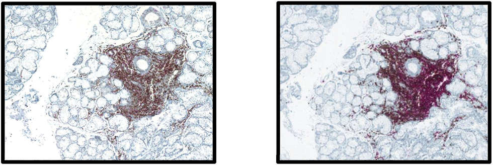
Results: 28 patients with sicca syndrome and MSGB with FS ≥1 were analyzed; 16 patients had SS (8 with polyautoimmunity) and 12 without SS, of which 5 had only sicca syndrome without defined disease and 7 had autoimmune rheumatic diseases other than SS. The presence of atrophy was significantly greater in autoimmune diseases other than SS and in SS cases with polyautoimmunity (p<0.05). All samples were positive for the 4 IHC biomarkers (Figure 1). When patients with SS and other autoimmune diseases were compared with respect to MSGB and IHC findings, the majority presented CD20:CD3 ratios ≤ 2:1 as well as CD4:CD8 ratios ≤ 2:1, with no statistically significant differences between the two groups (Table 1).
Conclusions: Histopathological and IHC findings were analyzed in Colombian MSGB patients with sicca symptoms, mainly diagnosed with SS. Regarding IHC, the ratios were mainly CD20:CD3 ≤ 2:1, as well as CD4:CD8 ≤ 2:1. A higher frequency of glandular atrophy was found in patients with SS and polyautoimmunity, as well as in patients with other autoimmune diseases. These findings are novel in the Latin American population and may serve to better define the subphenotypes according to biopsy.
Immunohistochemistry. (1a left) TCD4+/TCD8+ lymphocytes determined by CD4:CD8 (CD4, brown; CD8, red). (1b right) TCD8+ lymphocytes determined by CD20:CD3 (CD20, red; CD3, brown)

Description between patients with SS and other autoimmune diseases in terms of BGSM and immunohistochemical findings.
| No ptes/
| Focus score | Inflamation | CD20:CD3 | CD4:CD8 | |||||
|---|---|---|---|---|---|---|---|---|---|
| 1 - 2 | ≥2,1 | Mild | Moderate | Severe | 2:1 | 3:1 – 4:1 | 1:1 - 2:1 | 3:1 – 4:1 | |
| 16
| 13 (81,3%) | 3 (18,8%) | 3 (18,8%) | 10 (62,5%) | 3 (18,8%) | 12 (75%) | 4
| 9 (56,3%) | 7 (43,8%) |
| 6
| 6 (100%) | - | - | 5 (83,3%) | - | 5 (83,3%) | 1 (16,7%) | 4 (66,7%) | 2 (33,3%) |
| 1
| - | 1 (100%) | - | 1 (100%) | - | 1 (100%) | - | 1 (100%) | - |
| 1
| 1 (100%) | - | - | 1 (100%) | - | 1 (100%) | - | 1 (100%) | - |
| 7
| 6 (85,7%) | 1 (14,3%) | - | 7 (100%) | - | 5 (71,4%) | 2 (28,6%) | 3 (42,9%) | 1 (57,1%) |
| 4
| 4 (100%) | - | 1 (25%) | 3 (75%) | - | 3 (75%) | 1 (25%) | 4 (100%) | - |
| 1
| 1 (100%) | - | - | 1 (100%) | - | 1 (100%) | - | 1 (100%) | - |
| 1
| 1 (100%) | - | - | 1 (100%) | - | - | 1 (100%) | 1 (100%) | - |
SS: Sjögren’s syndrome, SLE: systemic lupus erythematosus, MCTD: mixed connective tissue disease, APS: antiphospholipid syndrome, RA: rheumatoid arthritis, SSc: systemic sclerosis, IIM: idiopathic inflammatory myopathy, PBC: primary biliary cholangitis. BGSM: minor salivary gland biopsy, IHQ: immunohistochemistry.
*Patients with polyautoimmunity.
(There were no statistically significant differences).
REFERENCES: NIL.
Acknowledgements: NIL.
Disclosure of Interests: None declared.