
Background: Current ultrasound (US) courses focus on the correct and accurate acquisition of images and the identification and scoring of abnormalities rather than how these imaging findings are used in clinical decision making. APLAR Academy considered a new approach to learning through the use of different clinical scenarios and pre- and post- test probability models. Point of care rheumatology ultrasound (POCRUS) as a sensitive and specific prediction tool has gained ground with EULAR recommendations for large vessel vasculitis (LVV) and polymyalgia rheumatica (PMR) criteria.
Objectives: Using a POCRUS approach, clinical cases were developed to illustrate how US findings might influence the management of 5 common clinical scenarios - monoarthralgia, oligoarthralgias, small joint polyarthralgias, polymyalgia, or LVV. The course aimed to illustrate and provide a forum for discussion on how POCRUS might facilitate patient management using a clinical, probability-based diagnostic algorithm with the intention of leading to faster and more accurate diagnoses, complementing traditional US teaching.
Methods: An online POCRUS course of six sessions was delivered (the first being an introduction to the concept), consisting of lectures on scenarios and case discussions where a novel probability-based model was used. Opportunities were provided for interactions on how POCRUS could be integrated into clinical practice as an adjunct. Pre- and post-course online surveys were undertaken to examine the current US landscape and whether the aims were met post course.
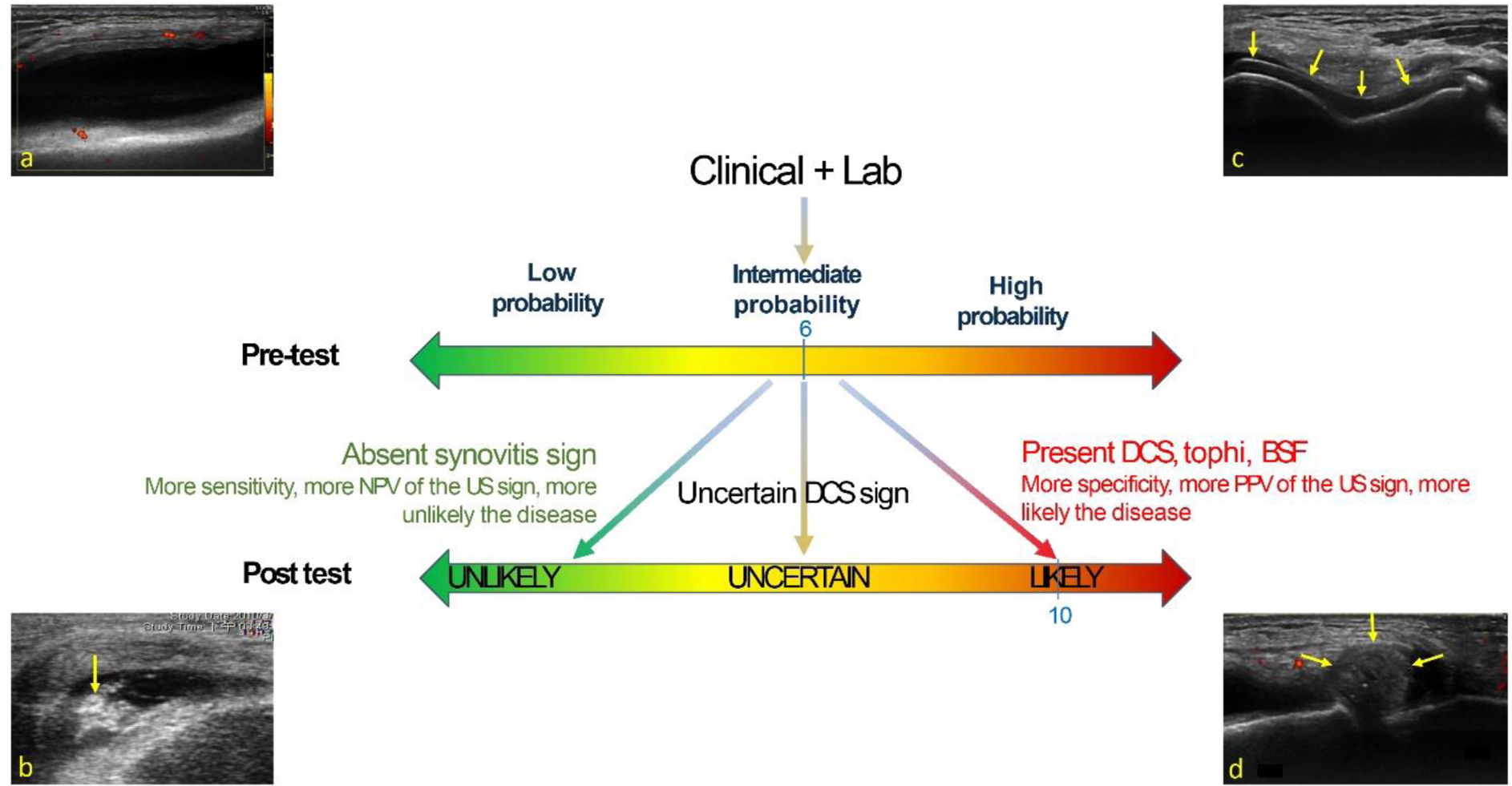
Results: Each clinical session consisted of a 45-minute lecture with rotating break-out group case discussions, on how POCRUS as an adjunct to clinical reasoning, can help transition from pre-test to post-test probability of target diagnoses (Figure 1).
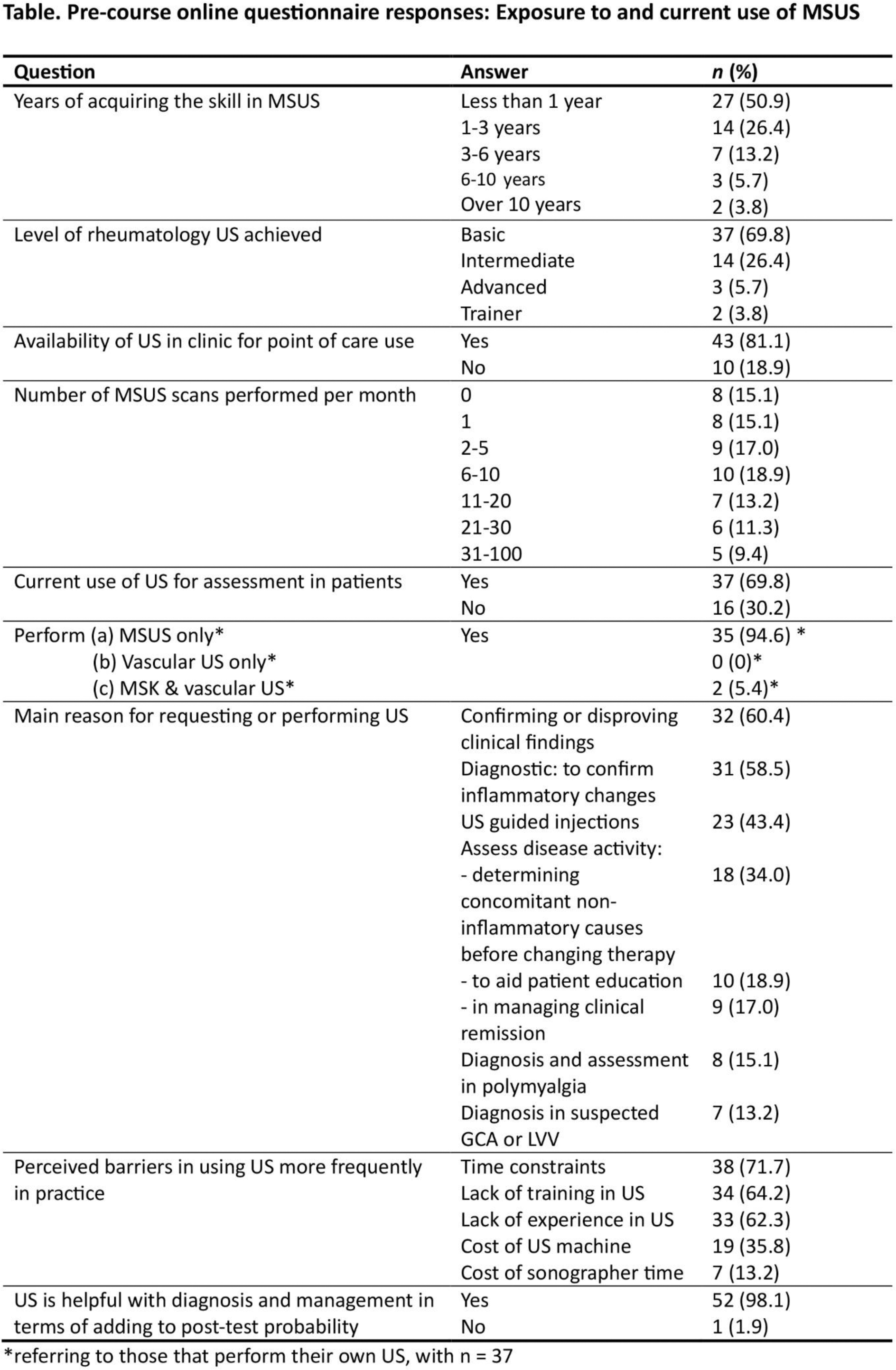
53/75 (70.7%) attendees completed a pre-course online questionnaire (Table 1). Despite POCRUS availability (81.1%), only 47.2% performed up to 5 scans/month. The main indication was musculoskeletal US (MSUS), with few performing vascular scans. Barriers included time constraints, and lack of training and experience. The vast majority (98.1%) reported that POCRUS may help with diagnosis and management of rheumatic disease by adding to post-test probability.
Post-session questionnaires revealed that 93-100% of attendees believed that US would fit well into and add to their clinical practice. The break-out sessions and case-based panel discussions were appreciated as opportunities to ask questions of the expert facilitators and allowed more focus on applying knowledge gained from the prior lecture into practice. 80-93% of attendees reported that their use of US in the subject covered would increase post course. In the vasculitis session 25% reported that the complex imaging procedure was beyond their skill set.
Proposed methods to improve learning outcomes and audience participation included pre-selection of participants into basic versus experienced learners. This will enable organizers to stratify the target audience based on US experience. In future, live demonstrations and hands-on teaching could be added.
Conclusion: The APLAR scenario-based online education in POCRUS was well received. Almost all reported US added to post-test diagnostic probability and disease management, adding to clinical confidence. POCRUS has potential implications in rheumatology practice for reducing subjective variations in clinical evaluation and diagnostic errors. This should facilitate one-stop consultations for early arthritis or fast-track clinics. POCRUS can represent a major future addition to traditional teaching in rheumatology and ultrasonography with potential in research for formulation and application of predictive diagnostic algorithms.
REFERENCES: NIL
POCRUS probability pathway for gout sequentially applying ACR EULAR gout criteria and sensitive (synovitis)/ specific (double contour, tophi, brightly stippled foci, erosions) ultrasound signs. (a) Synovitis (b) Brightly stippled foci (c) Double contour sign (d) Tophi


Acknowledgements: We would like to acknowledge the APLAR Academy, APLAR Education Committee and APLAR Secretariat for providing the funding, opportunity, and necessary support to organise and report this Webinar series. We would like to thank Professor Debashish Danda, President of APLAR, and Professor Lai Shan Tam, Convenor APLAR Academy, for their encouragement and support for this course.
Disclosure of Interests: None declared.