
Background: Synovial biopsies are conducted for clinical or research purposes. Their main aim in the clinical setting is assisting in arthritis diagnosis, complementing the synovial fluid analysis and other diagnostic tests, in particular if infections, synovial tumors, granulomatous or deposition diseases are suspected. The indication for synovial biopsies and the established diagnosis can be quite diverse across both medical and surgical departments.
Objectives: To compare the histopathological diagnosis of synovial specimens of biopsies with origin in the Rheumatology Department versus other Departments, in a Portuguese tertiary Center; To increase awareness for non-rheumatic synovitis etiologies in the rheumatology community.
Methods: This was a single center, cross sectional study of all synovial specimens received at the Pathology Department in Hospital de Santa Maria (Lisboa, Portugal), between 2010 and 2022. Histological diagnoses were classified as inflammatory or non-inflammatory lesions. Inflammatory lesions were further divided into non-granulomatous (low-grade or high-grade synovitis according to Krenn’s Score and infectious arthritis; low-grade synovitis also included cases of post-traumatic and mechanical causes), granulomatous, and crystal or foreign-body induced. Non-inflammatory lesions were subdivided into tumor-like and others. The categories were then matched for comparison according to its origin’s Department (Rheumatology versus Orthopedics, Plastic Surgery, General Surgery, Stomatology, Internal Medicine, Infectious Diseases and Pediatrics as a group).
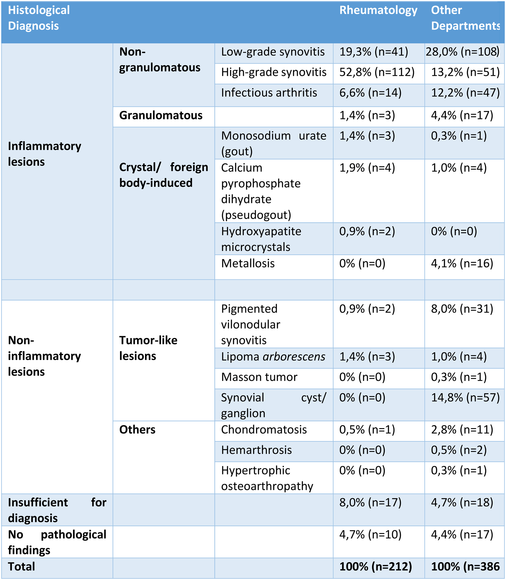
Results: A total of 598 reports pertaining to 539 patients (306 female, 56.8%) with an average age of 50.2 years old (ranging from 0 to 93) were reviewed. 212 specimens (35.4%) were from the Rheumatology Department while 386 (64.5%) from other Departments. Most of the cases from Rheumatology were classified as high-grade synovitis (n=112, 52.8%), whereas the largest group of the cases from other Departments were low-grade synovitis (n=108, 28.0%). The Rheumatology Department also had more cases of crystal induced diseases (4.2% vs 1.3%). Other Departments had a higher percentage of septic arthritis (12.2% vs 6.6%), granulomatous lesions (4.4% vs 1.4%), metallosis (4.1% vs 0%), tumor-like lesions (24.1% vs 2.4%) and chondromatosis (2.8% vs 0.5%). In the Rheumatology cases, after laboratory and histological results, there was a change in diagnosis in 61.8% (n=131) of the cases, mainly due to exclusion/ confirmation of septic arthritis and/or crystal-induced arthritis.
Conclusion: Synovial histopathological evaluation can be determinant for the diagnosis of a broad range of synovitis etiologies. As expected, most of the cases referred from the Rheumatology Department were high grade inflammatory lesions, whereas cases suggestive of other diagnosis such septic arthritis, tumor-like lesions or mechanical causes are mainly referred from other specialties. Taking in consideration that synovitis is a common manifestation of inflammatory and non-inflammatory diseases, the recognition of the relevance of integrating histological data in the diagnosis workup of synovitis is fundamental for a more precise medicine in the Rheumatology practice.
REFERENCES: NIL.
Table 1. Histological diagnosis distribution according to the Department of origin (Rheumatology versus other Departments).

N = number
Acknowledgements: NIL.
Disclosure of Interests: None declared.