fetching data ... 
Background: Rheumatoid arthritis (RA) is an autoimmune disease that causes destructive arthritis. In recent studies using single-cell analysis, the importance of clonally expanded cytotoxic T cells in the pathogenesis of this disease has been reported.[1,2] On the other hand, RA exhibits heterogeneity such as age of onset, seropositivity of autoantibodies, and extra-articular involvements, with varying prognoses and treatment responses. Therefore, we hypothesized that the role of the cytotoxic T cells in the immunological pathogenesis may differ according to clinical characteristics.
Objectives: To examine the involvement of CX3CR1-positive cytotoxic T cells which specifically produce granzyme B and perforin in the pathogenesis of RA, stratified by clinical characteristics.
Methods: Fresh peripheral blood was obtained from 78 treatment-naïve, active RA patients, 9 patients with difficult-to-treat RA, and 16 healthy individuals. Using flow cytometry, the proportions of CX3CR1-positive CD4 T cells and CX3CR1-positive CD8 T cells in peripheral blood were measured. The proportion of cytotoxic T cell subsets and their correlation with disease activity were compared between patients stratified based on characteristics. The infiltration of CX3CR1-positive CD4 T cells and CX3CR1-positive CD8 T cells into the affected synovial tissues was assessed using immunohistochemical staining of synovial biopsy samples from RA patients.
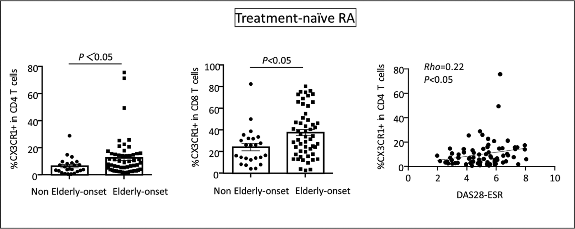
Results: CX3CR1-positive CD4 T cells in peripheral blood from patients with treatment-naïve RA were significantly increased compared to healthy individuals, while no significant difference was observed in CX3CR1-positive CD8 T cells. However, in elderly-onset RA patients (EORA), both CX3CR1-positive CD4 T cells and CX3CR1-positive CD8 T cells were significantly increased compared to non-EORA patients. Correlation analysis with disease activity indices showed positive correlation between the proportion of CX3CR1-positive CD4 T cells and CX3CR1-positive CD8 T cells and DAS28-ESR. In difficult-to-treat RA cases, the increase in CX3CR1-positive CD4 T cells and CX3CR1-positive CD8 T cells was also observed despite a history of multiple biologic agent treatment. Immunohistochemical analysis of synovial biopsy samples from EORA patients revealed massive infiltrations of CX3CR1-positive CD4 T cells and CX3CR1-positive CD8 T cells.
Conclusion: Our study suggests that the immunopathogenesis of RA varies according to the age of onset, with CX3CR1-positive cytotoxic T cells playing a role in EORA by contributing to disease activity and local synovial inflammation. Furthermore, CX3CR1-positive CD4 and CD8 cytotoxic T cells may be relevant for treatment resistance of RA.
REFERENCES: [1] Moon JS, et al. Nat Commun. 2023 Jan 19;14(1):319.
[2] Argyriou A, et al. Nat Commun. 2022 Jul 13;13(1):4046.
Acknowledgements: NIL.
Disclosure of Interests: None declared.