fetching data ... 
Background: B-lines (BLs) and pleural line alterations (PLA) are the typical lung ultrasound (LUS) findings for detection and severity assessment of systemic sclerosis-associated interstitial lung disease (SSc-ILD). Recently, LUS scores have shown to correlate with the extent of ILD and radiological abnormalities assessed by automated quantitative computed tomography (qCT). Nevertheless, one of the limitations of LUS remains the possibility of exploring only the superficial areas of the lung parenchyma.
Objectives: The aim of the study is to compare quantitative LUS scores with 3-levels qTC analysis (apices, midfields and lung bases) and to evaluate their possible association with ILD extension of lung surface and core.
Methods: During the period 2021-2023, consecutive SSc patients underwent on the same day CT and LUS, performed by two blinded certified operators using the 14 intercostal space assessment technique. The total number of BLs was collected, and the quantitative PLA score proposed by our centre was applied, dividing each lung into 3 levels. CT images were assessed by two thoracic radiologists for ILD definition and then analysed via automated texture analysis software, quantifying the volumes of total ILD, ground-glass (GG) and reticulations (RET), also differentiating ILD volume of the lung surface and core.
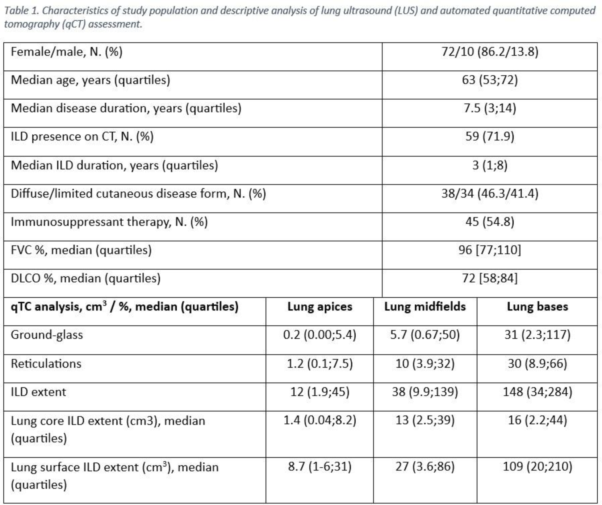
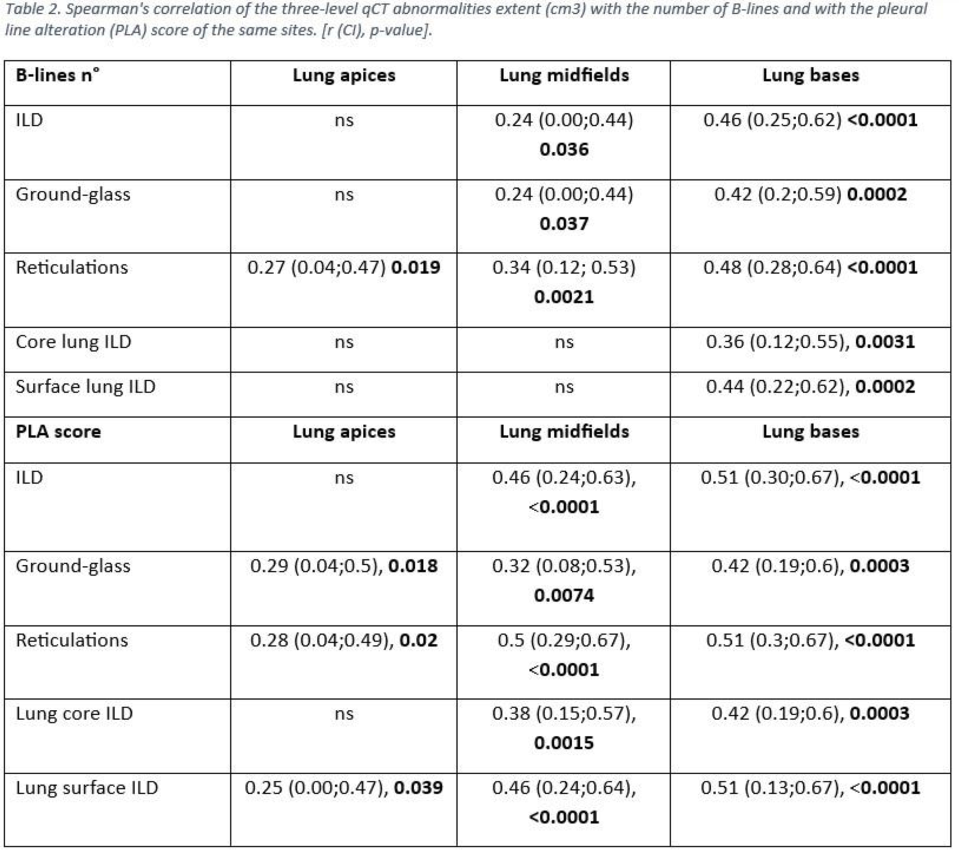
Results: Eighty-two patients were enrolled, with 59 (72%) presenting ILD on CT (Table 1). Median number (IQR) of B-lines was 1 (0;4) for apices, 6.5 (1;16) for mid-fields and 11 (3;26) for lung bases. Median PLA score of the apical, middle and basal lung fields was 1 (0;2), 5 (2;8) and 6 (2;11), respectively. Detailed qCT analysis is reported in Table 1. Both BLs number and PLA score of mid-basal lung fields correlated with the extent of GG, RET and ILD of the same level at qCT (p<0.005). The PLA score of lung apices correlated with the extent of GG and reticulations of the same site on qCT, whereas the number of BLs correlated only with reticulations (p<0.05). The extent of ILD, ground-glass and reticulations of the lung surface on qCT was significantly higher than the equivalent of the lung core (p<0.05). The basal lung BLs number was found to correlate with superficial and core ILD extent (p<0.005). The PLA score of the apices, mid-fields and bases correlated with surface lung ILD, GG and reticulations (p<0.05), and the mid-basal PLA score also correlated with deep lung alterations on qCT (p<0.02) (Table 2).
Conclusion: Our study confirms and expands on the recently emerged findings on the correlation of LUS with ILD extent at qCT, suggesting the ability of LUS to reflect lung morpho-structural changes, especially at basal lung, most affected by SSc-ILD. For the first time, LUS (particularly the PLA score proposed by our centre) was found to be associated with ILD volume of the lung core. This is probably because structural changes in the lung core would be reflected in the lung surface, where SSc-ILD is prevalent. These results may suggest the possibility of overcoming the traditional LUS limitation of investigating only the superficial lung parenchyma.
REFERENCES: [1] Gutierrez M et al. Radiol Med, 2019.
[2] Xie, H.Q et al. Arthritis Res Ther, 2019.
[3] Radić M. et al. Diagnostics (Basel), 2023.
[4] Bruni C et al. Diagnostics (Basel), 2022.
[5] Mohammad Reza Beigi D et al. Rheumatol Oxf, 2023.
Acknowledgements: NIL.
Disclosure of Interests: None declared.