fetching data ... 
Background: Scleroderma or systemic sclerosis (SSc) is a rare multisystemic autoimmune disease. It initiates with endothelial damage and inflammatory infiltrate, leading to fibrosis in various organs. Activation of different immune cells induces the transformation of endothelial cells and fibroblasts into myofibroblasts, responsible for excessive production of extracellular matrix components and reactive oxygen species. A better understanding of the pathophysiology leading to fibrosis and tissue damage is necessary to identify therapeutic targets. CEMIP (for “Cell migration-inducing protein” also called KIAA1199 and Hybid for “Hyaluronan-binding protein”) binds to hyaluronic acid (HA) and mediates its depolymerization into low molecular weight HA, leading to inflammation[1]. CEMIP expression is increased in cancer cells and positively regulates epithelial-mesenchymal transition, cell survival, growth and invasion. This work is based on our previous research underscoring a pro-inflammatory and a pro-fibrotic role of CEMIP in osteoarthritis (OA). Overexpression of CEMIP in OA tissues (cartilage and synovial membrane) is linked to the inflammatory environment. Moreover, CEMIP regulates pathways involved in OA and fibrosis such as Wnt/β-catenin and TGF-β[2,3].
Objectives: In the present study, in viv o CEMIP expression is investigated in the skin of HOCL-induced SSc mouse model compared to healthy skin. In vitro regulation of inflammation and fibrosis using human dermal fibroblasts (HDF) by CEMIP is also studied.
Methods: For in vivo study, SSc mouse model was obtained by sub-cutaneous daily injection of hypochlorous acid (HOCl). Mice were sacrificed after 7, 14, 28 and 42 days of HOCl injections. CEMIP mRNA expression level was assessed in mouse skin. CEMIP protein expression was also investigated in vitro using HDF subjected to TGF-βand IL1-β stimulation. Inflammatory and fibrotic markers levels were then evaluated after CEMIP silencing (using 2 specific shRNA).
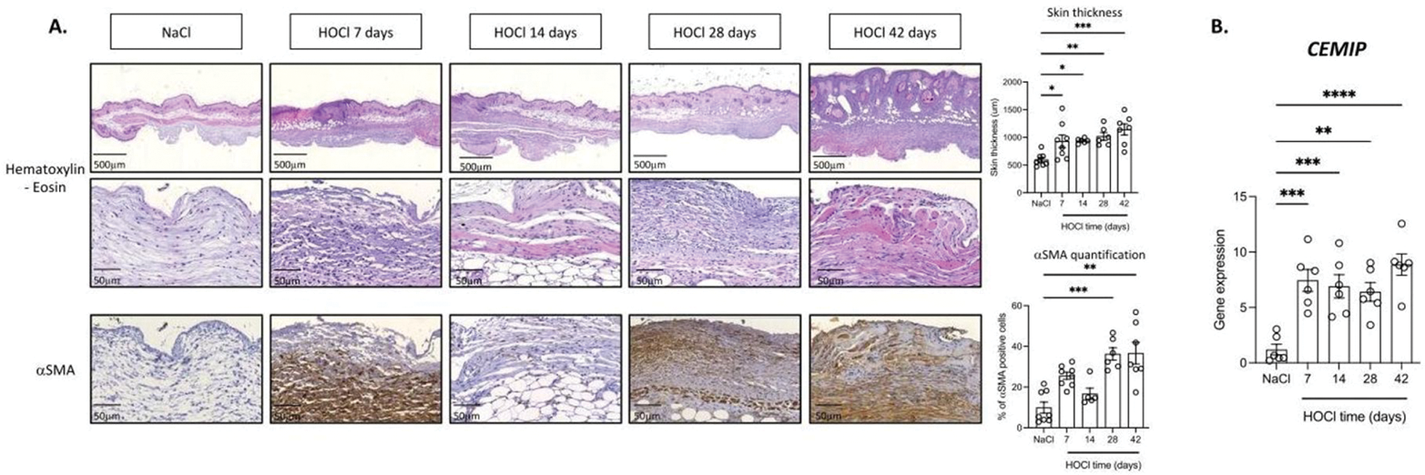
Results: As expected, skin thickening and fibrosis (αSMA expression) were observed in the skin of HOCL-injected mice compared to healthy skin (Figure 1A). Similarly, CEMIP mRNA expression was significantly overexpressed in the skin of these mice (Figure 1B). Moreover, its expression was positively correlated with inflammatory markers (IL6: r = 0.60, p = 0.0002; IL1B: r = 0.53, p = 0.001), fibrotic markers (αSMA: r = 0.42, p = 0.015; FN1: r = 0.39, p = 0.026; COL1A1: r = 0.51, p = 0.002; COL3A1: r = 0.45, p = 0.008) and skin thickness (r = 0.54, p = 0.001). In vitro experiments were performed on HDF stimulated with IL1-βand TGF-β. CEMIP protein level was significantly increased after 1 day of IL1-β stimulation (p = 0.031) and after 7 days of TGF-β stimulation (p = 0.031). Moreover, CEMIP silencing triggered decreased secretion of IL6 induced by IL1-β stimulation and decreased expression of αSMA induced by TGF-β stimulation.
Conclusion: CEMIP was overexpressed in the skin of HOCl-induced SSc mice and positively correlated with SSc features (skin thickness, markers of inflammation and fibrosis). Its expression is increased in inflammatory and fibrotic conditions in HDF. Finally, CEMIP regulated inflammatory and fibrotic markers.
REFERENCES: [1] Yoshida, H. et al. KIAA1199, a deafness gene of unknown function, is a new hyaluronan binding protein involved in hyaluronan depolymerization. Proc Natl Acad Sci U S A 110 , 5612–5617 (2013).
[2] Deroyer, C. et al. CEMIP (KIAA1199) induces a fibrosis-like process in osteoarthritic chondrocytes. Cell Death Dis 10 , (2019).
[3] Deroyer, C. et al. CEMIP (KIAA1199) regulates inflammation, hyperplasia and fibrosis in osteoarthritis synovial membrane. Cellular and Molecular Life Sciences 79 , (2022).
Representative histological pictures of mouse skin sections after 0, 7, 14, 28 and 42 days of HOCl injection stained with hematoxilin-eosin and aSMA antibody. Skin thickness was measured and expressed in µm and aSMA expression was quantified and expressed in percentage of positive stained cells ( A ). RT-qPCR analysis of CEMIP gene in skin of HOCl induced mice (0, 14 and 42 days of injection) ( B ).
Acknowledgements: NIL.
Disclosure of Interests: None declared.