fetching data ... 
Background: Mast cells (MCs), tissue-resident granulocytes best known for their involvement in allergic reactions, also play pivotal roles in fibrosis, immune modulation, and angiogenesis, all key components of systemic sclerosis (SSc) pathology. Previous studies with limited samples have highlighted the increased MC numbers and degranulation in SSc, yet the implications of these changes for disease pathophysiology and clinical manifestations remain unclear. Furthermore, the precise signaling pathways linking MCs to SSc pathogenesis are not fully understood.
Objectives: This study aims to better understand the role of MCs in SSc pathogenesis and clinical stratification, with a focus on their contribution to vasculopathy.
Methods: We conducted single-cell RNA sequencing (scRNAseq) on skin biopsies (SSc=5, control =2) using the BD Rhapsody platform, which is optimized for granulocyte capture and particularly suitable for studying these cells. Spatial transcriptomics was performed using Visium HD for single-cell-scale resolution. Toluidine blue staining was employed to assess MC density and degranulation in skin samples, and clinical correlations were explored in a cohort of SSc patients (n=93).
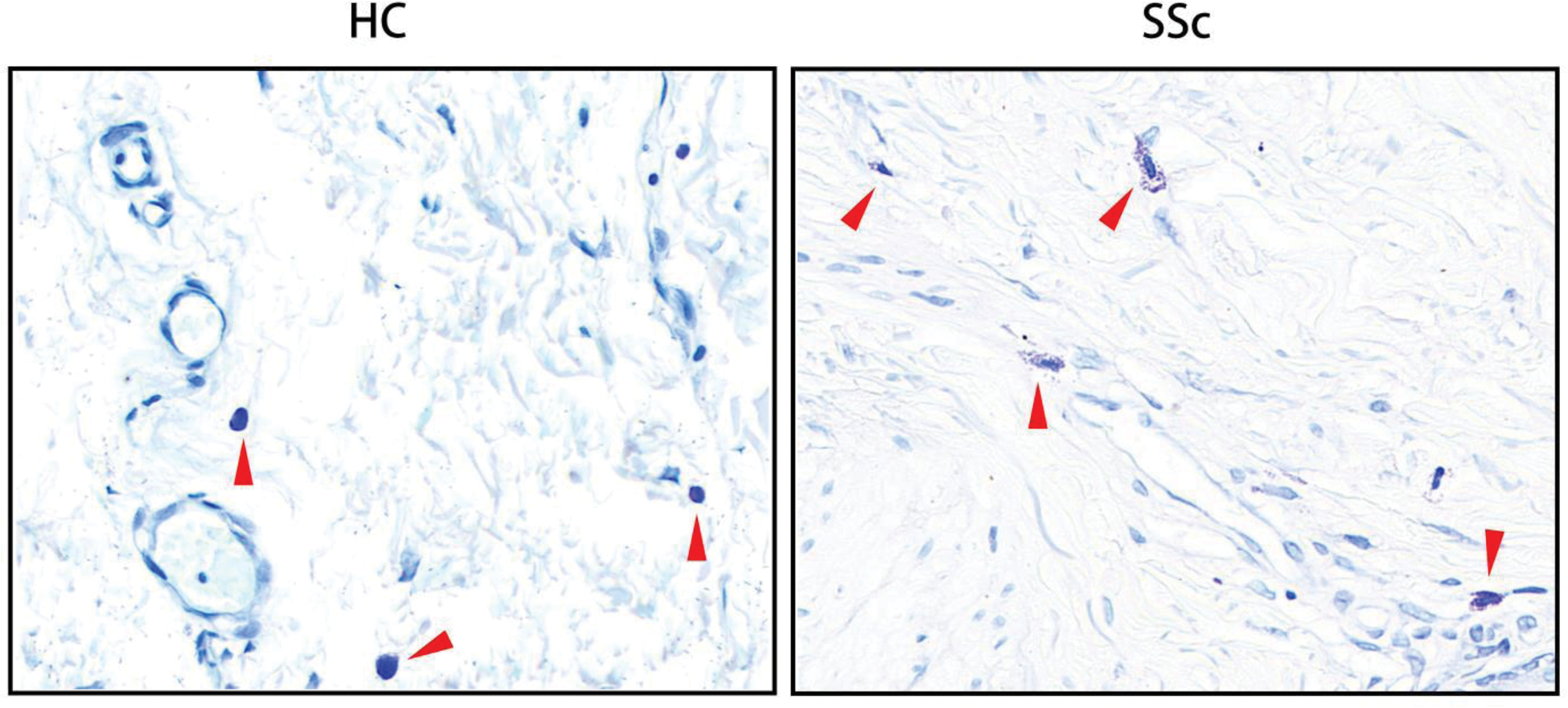
Results: MC density was significantly increased in SSc skin. Functional analysis indicated enhanced MC activation in SSc. Cellchat analysis revealed increased crosstalk between MCs and endothelial cells in SSc. Notably, LIF/LIFR signaling between MCs and endothelial cells was significantly enriched in SSc, with MCs identified as the primary source of LIF. MCs in SSc, particularly in diffuse cutaneous SSc (dcSSc), exhibited elevated LIF expression. For spatial transcriptome data, neighborhood enrichment analysis by Squidpy revealed closer proximity between MCs and endothelial cells. Cellular communication between MCs and endothelial cells via the LIF-LIFR axis was visualized in Visium HD data. Toluidine blue staining confirmed increased MC numbers and degranulation in SSc skin compared to healthy controls. Importantly, patients with digital ulcers (DU) or pruritus exhibited higher densities of degranulated MCs and more pronounced MC degranulation.
Conclusion: MCs are enriched and activated in SSc skin, contributing to vasculopathy through mechanisms involving LIF/LIFR signaling. These findings highlight the potential of MCs as therapeutic targets in SSc.
REFERENCES: NIL.
Representative images of mast cell infiltration and degranulation stained with toluidine blue in skin biopsies (mast cells were stained blue-purple). Left: Resting mast cell (arrowheads) in HC skin. Right: Degranulated mast cells (arrowheads) in the proximaty of blood vessels in SSc skin.
Acknowledgements: NIL.
Disclosure of Interests: None declared.
© The Authors 2025. This abstract is an open access article published in Annals of Rheumatic Diseases under the CC BY-NC-ND license (