fetching data ... 
Background: Sjögren’s syndrome (SS) is a prototypical chronic autoimmune disease characterized by lymphocytic infiltration of the exocrine glands, especially the salivary and lacrimal glands. Glandular tissue degradation is one of the pathologic steps in SS, and MMP-9 is an important determinant of the destruction of the basement membrane through the degradation of gelatins and type IV collagen. MMP-9 expression is significantly higher in minor salivary glands and saliva of SS patients compared to healthy controls, and is increased in tissue samples from SS patients in a manner that correlated with the disease severity. Syndecan-4 (SDC4) is a ubiquitously expressed transmembrane heparan sulfate (HS) proteoglycan composed of an extracellular domain, a signature transmembrane and a cytoplasmic domain. It regulates interactions with the extracellular matrix and is transiently upregulated in conjunction with integrins during tissue repair. Additionally, mice lacking SDC-4 show delayed wound healing and impaired angiogenesis. SDC-4 can be proteolytically released from the cell surface through ectodomain shedding by sheddase. MMP-9 is well recognized as one of the important sheddase of SDC-4. So, SDC-4 shedding might be related with tissue inflammation in SS due to the increased expression of MMP-9; however, the relationship between SDC-4 shedding and MMP-9 in SS has not been reported to date.
Objectives: We aimed to investigate the relationship between SDC-4 shedding and MMP-9 in blood and inflamed salivary gland tissues in SS.
Methods: We enrolled 70 patients with SS and 35 controls in this study. For animal experiments, female NOD/ShiLtJ between 6 and 12 weeks of age and sex-, and age-matched C57BL10 mice were used. The concentrations of SDC-4 MMP-9 were analyzed using western blotting. The severity of inflammation of the minor salivary gland from patients and submandibular gland (SMG) tissues from mice was assessed by focus score (FS) and ration index ([area of inflammation/total glandular area of the section] × 100), respectively. For the HS treatment experiment, NOD/ShiLtJ mice aged 6–10 weeks were treated with 5 mg/kg HS intraperitoneally thrice per week, and the therapeutic effects on SMG tissues were evaluated at the end of 10 weeks of age.
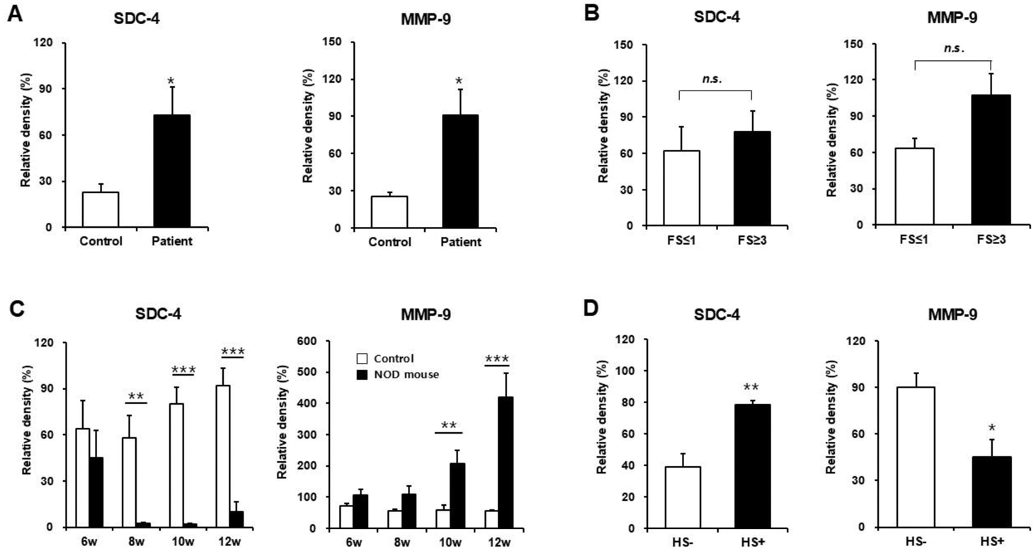
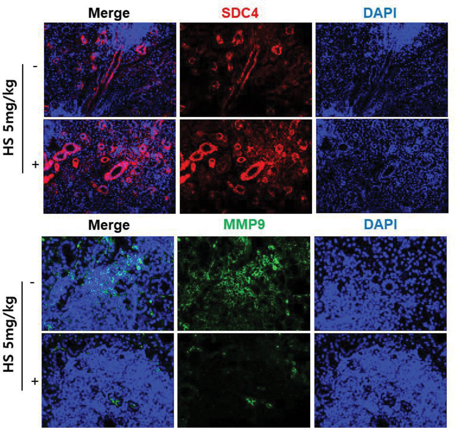
Results: Blood concentrations of SDC-4 (P = 0.029) and MMP-9 (P = 0.014) in patients with SS were statistically significant increased compared heathy controls (Figure 1A). There was also a trend toward increased blood concentrations of SDC-4 and MMP-9 in patients with a FS of 3 or higher than in patients with a FS of 1 or lower (Figure 1B). In the NOD/ShiLtJ mice, the expression of SDC-4 in the SMG tissues was significantly reduced compared to control mice starting at 8 weeks of age. The expression of MMP-9 was significantly increased in the SMG tissues of NOD/ShiLtJ mice at 10 and 12 weeks of age compared to controls. The concentration of MMP-9 in the SMG tissues of NOD/ShiLtJ mice showed an increasing trend with increasing inflammation, with a statistically significant difference at 12 weeks of age (P = 0.038). Additionally, increased SMG tissue inflammation was associated with increased blood concentrations of SDC-4 and MMP-9 (Figure 1C). The HS-treated group showed a significant decrease in the SMG tissue inflammation (P = 0.002), and B- and T-cell infiltration compared to the control mice. Additionally, a significant increase in SDC-4 concentrations (P = 0.004) and a decrease in MMP-9 concentrations (P = 0.021; Figure 1D & Figure 2) in the SMG tissues were observed in the HS-treated group. The concentrations of MMP-9 in the blood was also significantly reduced in the HS-treated group (P = 0.037).
Expression of syndecan-4 (SDC-4) and MMP-9 in blood and minor salivary gland tissues of patients with Sjögren’s syndrome (A & B) and blood and submandibular gland (SMG) tissues from NOD/ShiLtJ mice (C & D). (A) Concentrations of SDC-4 and MMP-9 in the blood from SS patients and healthy controls. (B) Comparison of SDC-4 and MMP-9 concentrations in blood according to the severity of minor salivary gland inflammation. (C) Concentrations of SDC-4 and MMP-9 in the SMG tissues from NOD/ShiLtJ mice. (D) Therapeutic efficacy of heparan sulfate (HS) treatment on expression of SDC-4 and MMP-9 in the SMG of NOD/ShiLtJ mice. n.s. = not significant; * = P < 0.05, ** = P < 0.05, *** = P < 0.001.
Representative immunofluorescence staining for syndecan-4 (red) and MMP-9 (green) in the tissue sections before and after heparan sulfate treatment. The sections were counterstained with 4′,6-diamidino-2-phenylindole (DAPI, blue).
Conclusion: These results suggest that the expression and activity of MMP-9, an SDC-4 sheddase, increased due to inflammatory response in salivary gland tissues, resulting in increased SDC-4 shedding. Additionally, the possibility of SDC-4 concentrations in blood as a biomarker reflecting the degree of salivary gland tissue inflammation may be considered.
REFERENCES: [1] Tzioufas, A.G.; Kapsogeorgou, E.K.; Moutsopoulos, H.M. Pathogenesis of Sjogren’s syndrome: What we know and what we should learn. J. Autoimmun. 2012, 39, 4–8.
[2] Bernfield, M.; Gotte, M.; Park, P.W.; Reizes, O.; Fitzgerald, M.L.; Lincecum, J.; Zako, M. Functions of cell surface heparan sulfate proteoglycans. Annu. Rev. Biochem. 1999, 68, 729–777.
[3] Ram, M.; Sherer, Y.; Shoenfeld, Y. Matrix metalloproteinase-9 and autoimmune diseases. J. Clin. Immunol. 2006, 26, 299–307.
Acknowledgements: NIL.
Disclosure of Interests: None declared.
© The Authors 2025. This abstract is an open access article published in Annals of Rheumatic Diseases under the CC BY-NC-ND license (