fetching data ... 
Background: Rheumatoid Arthritis (RA) is a chronic autoimmune disorder marked by joint inflammation and damage. Its heterogeneous presentation and lack of reliable biomarkers often necessitate a trial-and-error approach in treatment. PET/CT, which combines the molecular sensitivity of PET with the anatomical images of CT, offers a powerful quantitative tool for early disease assessment in the whole body and treatment monitoring. Developing novel PET tracers targeting RA-specific pathways, such as cytokines and immune cells, holds promise for personalized care by stratifying patients based on immunological profiles and predicting treatment responses.
Objectives: This study investigates the potential of targeting the pro-inflammatory cytokine tumour necrosis factor (TNF) using a small engineered biomolecule called an affibody (Z1085) as a PET tracer.
Methods: TNF availability and distribution were assessed by measuring its concentration in synovial fluid and plasma from RA patients using Luminex immunoassay. Immunofluorescence (IF) staining of RA synovial tissue (ST) was performed using a commercially available TNF antibody in a cohort of the three synovial histological pathotypes: (1) fibroid, (2) diffuse-myeloid, (3) lymphoid-myeloid. Binding affinity of the radiolabelled tracer Z1085-DOTA-[68Ga] was determined using surface plasmon resonance (SPR) in competition with the anti-TNF agent etanercept. A preclinical in vivo biodistribution study in an Antigen-Induced Arthritis (AIA) rat model was conducted to determine the optimal tracer concentration: 1μg/kg (n=4), 5μg/kg (n=4) or 25μg/kg (n=5). Tracer uptake (%ID/gram) was calculated at 30 minutes post-injection (p.i.) for each organ. One dynamic μPET/CT scan per concentration was performed for 1h p.i. to investigate the imaging properties and targeting of TNF affibody. The outcome of the PET/CT images was interpreted visually and quantitatively by calculating the mean of the standardized uptake value (SUV mean ).
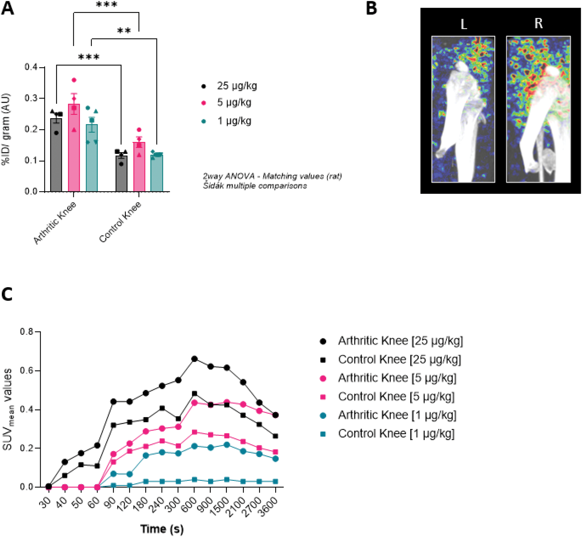
Results: TNF levels were significantly higher in synovial fluid than in plasma (p<0.05 ), suggesting accumulation in the joint, which is critical for achieving a high signal-to-background ratio of arthritic joints with PET imaging. IF staining of ST confirmed presence of TNF in inflamed joints in the diffuse-myeloid and lymphoid-myeloid pathotypes characterised by macrophage infiltration. The tracer exhibited a high binding affinity towards TNF (K D =1.1 nM), which was partly blocked by etanercept. Biodistribution studies revealed around two-fold increased uptake of [68Ga]-DOTA-Z1085 in inflamed knees compared to controls for each tracer concentration cohort ( p<0.01 and p<0.001 for 1 and 5-25 μg/kg, respectively), which correlated with the PET/CT images (Figure 1A-B). The SUV mean values seemed to decrease after 30 p.i. (Figure 1C). The principal route of excretion was through the urinary tract, accompanied by significant tracer accumulation in immune-associated organs (liver and spleen), which may be partly attributed to the metabolic clearance pathway of the radiotracer and partly to TNF overexpression resulting from systemic AIA-induced inflammation.
Conclusion: This study demonstrates the potential of [68Ga]-DOTA-Z1085 as a PET tracer for diagnostic use in RA. The tracer’s high binding affinity, favourable biodistribution with accumulation in inflamed joints highlight its promise for imaging of TNF in RA. However, histological validation and/or quantitative binding studies with a blocking compound are essential to assess systemic inflammation, tracer clearance and specificity. These findings lay the groundwork for further development and clinical application of this tracer in RA diagnosis and treatment monitoring.
REFERENCES: NIL.
A: Uptake of the radiolabelled TNF affibody [68Ga]-DOTA-Z1085 in arthritic knees compared to control knees in AIA rats at 30 minutes post-injection. Three tracer concentrations (1, 5 and 25 μg/kg) were tested in cohorts of 4-5 rats. B: PET image (3D view) of the right knee (R=inflamed) and left knee (L=control) of an AIA rat acquired over 1h using Z1085-DOTA-[68Ga] (5 μg/kg). C: SUV mean analysis of the dynamic PET scans of the AIA rat knees for each tracer concentration (1, 5 and 25 μg/kg) (n=1). The PET images were analyzed using vivoQuant software and the synovium region was segmented from the surrounding bones for a more accurate SUV mean measurement.
Acknowledgements: NIL.
Disclosure of Interests: None declared.
© The Authors 2025. This abstract is an open access article published in Annals of Rheumatic Diseases under the CC BY-NC-ND license (