fetching data ... 
Background: The ligamentum capitis femoris (LCF) is an important hip joint structure. Along with the articular cartilage, the LCF also undergoes a structural change in an injured or inflamed joint (Mande et al., 2003; Dehao et al., 2015; Sampatchalit et al., 2017). Its normal structure, particularly lectin histochemistry, remains insufficiently studied.
Objectives: This study aimed to examine the distribution of lectin receptors in the normal LCF during the postnatal period.
Methods: The hip joint of intact Wistar rats was the object of the study. Animals were euthanized on the first, seventh, 14th, 30th, 60th, and 90th days (n = 6; 36 rats total). Bouin-fixed and paraffin-embedded samples were stained with lectins conjugated with horseradish peroxidase (HRP): Peanut (PNA), Soybean (SBA), Helix pomatia (HPA), Perca fluviatilis (PFA), Wheat germ (WGA), Vicia sativa (VSA), Lens culinaris (LCA), Sambucus nigra (SNA). The intensity of staining was analyzed semiquantitatively.
Results: Throughout the follow-up period, the synovial lining cells covering the LCF stained very weakly (0/+); the modified basement membrane of the synovial intima showed a robust expression (from +++ to ++++); the cell nuclei showed a different reaction (from middle ++ to strong +++) to all studied lectins.
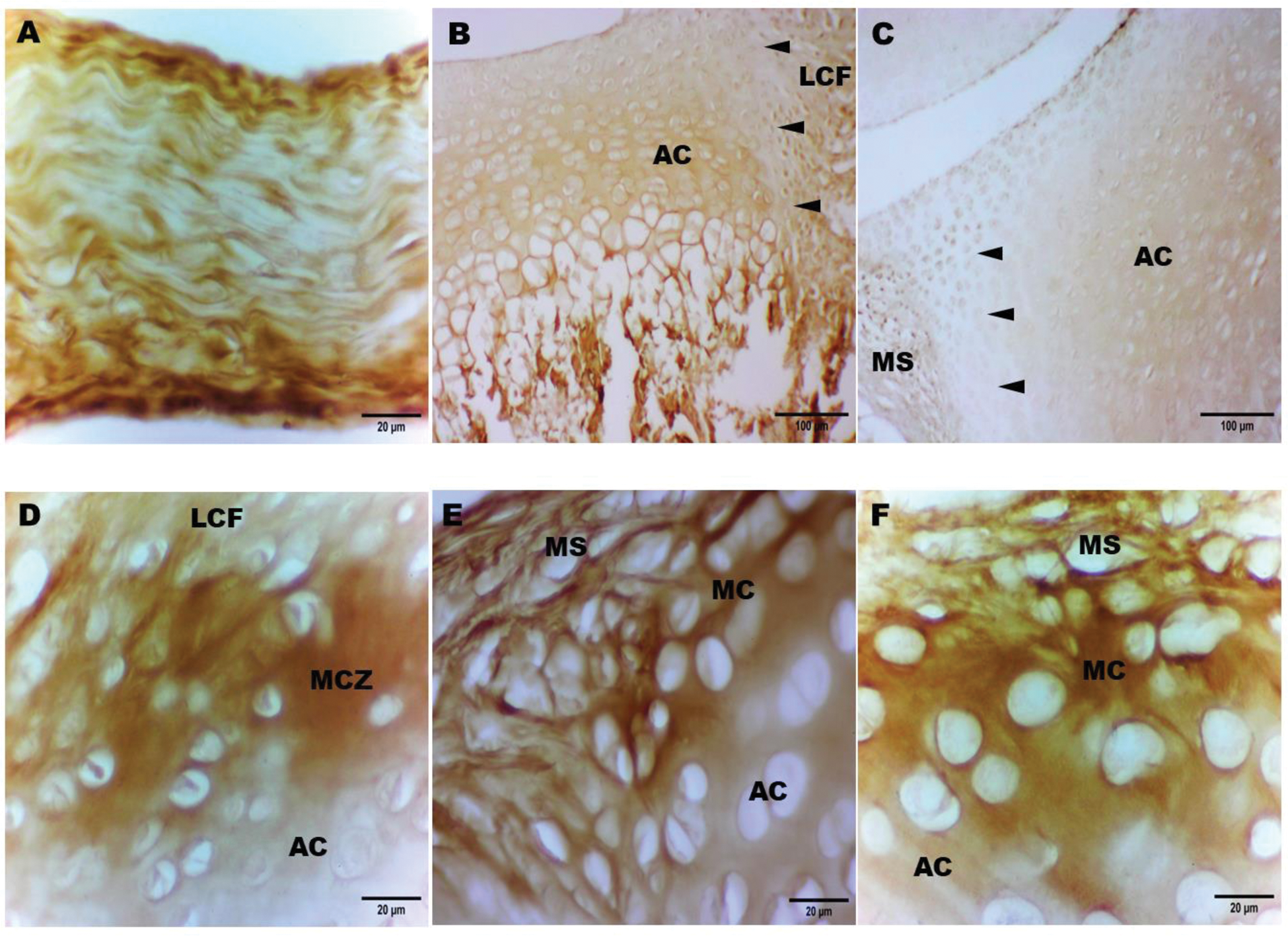
Till the 90th day, the extracellular matrix of the LCF showed a minimal (0/+) level of the SNA and a weak (+) expression of the PFA. The LCF fibers were stained very weakly (0/+) with the WGA till the seventh day and showed a weak (+) affinity from the 14th day. The mannose-specific lectins (LCA and VSA) revealed a very weak reaction (0/+) in the LCF fibers in newborns, weak (+) reaction till the 14th day, moderate expression (from + to ++) on the 30th day, and strong expression (+++) from the 60th day. The galactose-specific lectins (PNA, HPA, and SBA) showed weak staining (+) in the LCF fibers till the seventh day, moderate reaction (++) on the 14th day, moderately pronounced (++/ +++ for the PNA and HPA) and strong (+++ for the SBA) reaction from the 30th day (Figure 1A). From the 30th day, strong stained (except for the SNA) strip-like fibers were observed in the LCF. The wall of vessels, especially intima and adventitia, always showed a strong expression (+++) to the studied lectins. Till the seventh day, in the marginal part of the articular cartilage (acetabulum and caput femoris, respectively) that is in direct contact with the LCF woven into it, the expression of studied lectins was not observed. This so-called marginal cartilaginous zone (MCZ) is visualized as a “pale area” compared to the adjacent articular cartilage with pronounced lectin expression (Figure 1B). Subsequently, there was a robust (from +++ to ++++) expression of receptors to the PNA and SBA (14-90th day), HPA (14-30th day), LCA and VSA (30-90th day) in the MCZ (Figure 1D). On the contrary, no reaction with the LCA and VSA on the 14th day; HPA on the 60-90th day; WGA, PFA, and SNA throughout the follow-up period in the MCZ was observed.
Conclusion: The distribution of lectin receptors in the LCF is similar to the joint capsule. The lectin histochemistry of the marginal cartilaginous zone (MCZ) directly contacting the LCF, in turn, is similar to the marginal cartilage of the hip joint marginal transitional zone. The phenomenon of “pale areas” and the subsequent hyperexpression of lectins at the point of attachment of the LCF to the articular cartilage, by analogy with the marginal cartilage (Figure 1C, E, F), apparently plays the role of an innate non-specific lectin-mediated immunobiological barrier that would prevent excessive invasion of the LCF into the cartilage and might be relevant in immunoinflammatory diseases of the hip joint.
A , The LCF on the 30th day, SBA-HRP staining, x40. B , The attachment of the LCF to the acetabulum on the seventh day, SBA-HRP staining, x40. The arrows indicate the marginal cartilaginous zone (MCZ), which, like the marginal cartilage (MC, Figure 1C), is observed as a “pale area” compared to the adjacent articular cartilage (AC). C , The caput femoris (above) and acetabulum (below) on the seventh day, SBA-HRP staining, x40. The arrows indicate the marginal cartilage (MC), which is in direct contact with the marginal synovium (MS) and lectinhistochemically different from the adjacent articular cartilage (AC). D , The attachment of the LCF to the caput femoris on the 30th day, SBA-HRP staining, x40. The marginal cartilaginous zone (MCZ) with robust expression of lectin receptors, similarly to the marginal cartilage (MC, Figure 1E, F). E and F , The marginal transitional zone of the caput femoris (E) and acetabulum (F) on the 30th day, SBA-HRP staining, x40. A strong lectin expression in the marginal cartilage (MC) could be seen.
REFERENCES: NIL.
Acknowledgements: NIL.
Disclosure of Interests: None declared.
© The Authors 2025. This abstract is an open access article published in Annals of Rheumatic Diseases under the CC BY-NC-ND license (