fetching data ... 
Background: Systemic scleroderma (SSc) is an autoimmune disorder with diverse clinical manifestations. The pulmonary system is one of the primary organs affected by this condition. Lung involvement in SSc is associated with a substantial decline in both patient survival and quality of life.
Objectives: Thus, our objective was to evaluate the potential beneficial effects of vitamin D3 (VD3) and α-tocopherol acetate (ATA) on the morphology of the lungs, with a particular focus on changes in alveolocytes II type (A-II), in the preclinical model of SSc.
Methods: The experimental animals were divided into three groups: a control group (CG) consisting of 20 animals, and two experimental groups (EG#1 and EG#2) with 25 animals each. All animals were mature Wistar-line laboratory rats, weighing between 180 and 220 grams. For systemic scleroderma (SSc) modeling, animals in EG#1 received sodium hypochlorite (NaClO) injections following a previously established protocol [1]. In contrast, rats in the CG were administered an isotonic solution using the same schedule. In EG#2, rats received intramuscular injections of α-tocopherol acetate (ATA) at a dose of 10 mg/100 g of body weight and vitamin D3 (VD3) at 1000 IU/100 g of body weight over a 3-week period during the second half of the experiment. All animals were euthanized humanely 8 weeks after the initiation of the experiment. The cellular ultrastructure was examined using a PEM-125K electron microscope (Selmi, Ukraine).
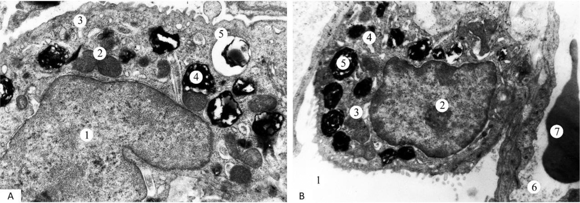
Results: Phenomena of hyperhydration were observed in A-II. The nuclei of the cells exhibited a fine-grained matrix (Figure 1A). In many A-II, chromatin granules were arranged along the inner surface of the nuclear membrane or clustered into separate aggregates (1). The perinuclear space was expanded. Mitochondria (2) were enlarged, with disoriented cristae. Alongside expanded Golgi apparatus (GA) components, fragmentation of rough endoplasmic reticulum (RER) membranes was evident (3). The number of ribosomes on these membranes was reduced. Disruptions in the submicroscopic structure were also found in the lamellar bodies (LB) (4, 5). Most LBs were located in the apical part of the cytoplasm of A-II. Many LBs exhibited irregular light spaces between the bimembranous osmiophilic lamellae. Some LBs are vacuolated with remnants of membranes. In addition, the cytoplasm of some cells contained homogeneous, medium-electron-density lipid-like inclusions. The apical surface of A-II appeared flattened due to a reduced number of microvilli. The basal membrane was of uneven thickness. After usage of VD3 and ATA, moderate pathological changes were still observed in A-II (Figure 1B). The cell nucleus (2) was characterized by the presence of fine-grained nucleoplasm and marginally located chromatin granules. The perinuclear space exhibited localized expansion. Mitochondria (3) of varying sizes and shapes were visualized, with some showing matrix clearing. The cisternae and tubules of the GA and RER were moderately expanded. The number of LBs (5) in the cytoplasm of A-II was increased. A significant number of microvilli, arranged in a mosaic pattern, were observed on the apical surface of the cells. The basal membrane exhibited localized thickening.
Conclusion: This study demonstrates the beneficial impact of vitamin D3 and α-tocopherol acetate on the ultrastructural organization of Type II alveolocytes in a experimentally induced model of systemic sclerosis.
REFERENCES: [1] Doskaliuk B, Zaiats L, Yatsyshyn R. AB0090 The biochemical basis of animal model of systemic sclerosis. Annals of the Rheumatic Diseases 2021;80:1074.
Acknowledgements: NIL.
Disclosure of Interests: None declared.
© The Authors 2025. This abstract is an open access article published in Annals of Rheumatic Diseases under the CC BY-NC-ND license (