fetching data ... 
Background: Antiphospholipid syndrome (APS) is an autoimmune disorder characterized by thrombosis, and/or pathological pregnancies, mainly recurrent abortion, due to the presence of persistently positive antiphospholipid antibody (aPL) [1]. Pregnancy loss, particularly in the first trimester, is a common manifestation of APS, but the pathogenic mechanisms involving trophoblast dysfunction are not fully explained [2]. Recent studies have demonstrated that aPL promotes senescence in human umbilical vein endothelial cells (HUVECs), contributing to pregnancy complications in APS mouse models [3]. Moreover, aPL has been implicated in accelerating the severity of Alzheimer’s disease, suggesting a potential link between APS and aging [4]. Despite these findings, the specific impact of aPL on trophoblast cell senescence and its role in APS-associated pregnancy loss remains unknown.
Objectives: To investigate the role of trophoblast cell senescence in APS-associated pregnancy loss and to explore its potential as a therapeutic target.
Methods: A total of 8 patients diagnosed with APS, characterized by high-titer aPL antibodies (>140 GPL U), thrombosis, and adverse pregnancy outcomes, were included in this study. For comparison, 16 healthy female volunteers with no history of adverse pregnancy, thrombosis, or autoimmune diseases were selected as controls. Normal human immunoglobulin G (NHIgG) and polyclonal APS-IgG antibodies were isolated using the NAb™ Protein G Spin Kit (Thermo Fisher Scientific). Villous tissue samples were collected from patients with APS and healthy controls during gestational weeks 6–10. Senescence levels in villous tissues from APS patients and trophoblasts treated with aPL antibodies were assessed using β-galactosidase staining. Western blotting (WB) was employed to examine the expression of senescence markers, and qPCR was used to evaluate changes in senescence-associated secretory phenotype (SASP) factors in trophoblast cells treated with APS-IgG.
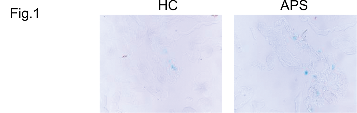
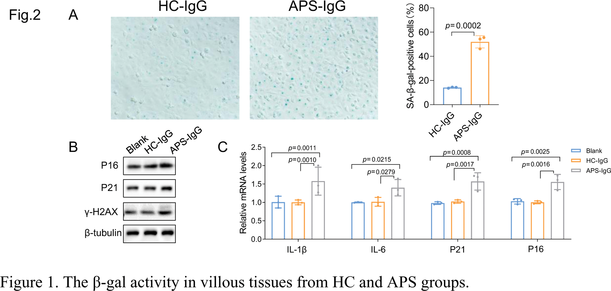
Results: SA-β-gal staining (positive β-galactosidase activity indicated by blue staining) revealed significantly higher SA-β-gal activity in villous tissues from the APS group compared to the healthy control (HC) group (Figure 1). To confirm that APS-IgG induced cellular senescence, IgG was isolated from the serum of APS patients and HC individuals and exposed to HTR-8/SVneo cells. SA-β-gal staining demonstrated an increased number of senescent cells in APS-IgG-treated trophoblasts compared to those treated with HC-IgG (Figure 2A). Additionally, APS-IgG elevated the protein and mRNA levels of senescence markers P16, P38, and γ-H2AX (Figure 2B-C). Considering that SASP factors are critical indicators of cellular senescence, we assessed changes in their mRNA levels. APS-IgG significantly upregulated the mRNA expression of key SASP factors, including IL-1β and IL-6.
Conclusion: High levels of cell senescence were observed in villous tissues from APS-associated pregnancy loss, and aPL was found to promote trophoblast cell senescence. These findings suggest a positive correlation between trophoblast senescence and APS. Targeting trophoblast cell senescence may provide a novel therapeutic strategy to improve adverse pregnancy outcomes in APS patients.
REFERENCES: [1] Knight JS, Branch DW, Ortel TL. Antiphospholipid syndrome: advances in diagnosis, pathogenesis, and management. BMJ 2023;380:e069717.
[2] Schreiber K, Sciascia S, de Groot PG, et al. Antiphospholipid syndrome. Nature Reviews Disease Primers 2018;4:17103.
[3] An R, Yang Y, Liu L, Li P. SAMD1 attenuates antiphospholipid syndrome-induced pregnancy complications. Immunity, inflammation and disease 2023;11:e1006.
[4] Katzav A, Faust-Socher A, Kvapil F, et al. Antiphospholipid syndrome induction exacerbates a transgenic Alzheimer disease model on a female background. Neurobiology of aging 2011;32:272-9.
The β-gal activity in villous tissues from HC and APS groups.
Effect of APS-IgG on trophoblast cell senescence. A. Representative images and quantification of SA-β-gal (+) trophoblast cells treated with HC-IgG or APS-IgG (100 µg/mL). B. The protein levels of P21, P16, and γ-H2AX in HTR-8/SVneo trophoblast cells treated with HC-IgG or APS-IgG (100 µg/mL). C. The mRNA levels of IL-1β, IL-6, P21, and P16 in HTR-8/SVneo trophoblast cells treated with IgG (100 µg/mL) from HC or APS groups.
Acknowledgements: NIL.
Disclosure of Interests: None declared.
© The Authors 2025. This abstract is an open access article published in Annals of Rheumatic Diseases under the CC BY-NC-ND license (