fetching data ... 
Background: Calcium-containing crystals are frequently observed in soft tissues that typically do not calcify, such as articular cartilage and peri-articular tissues, particularly in individuals with advanced osteoarthritis (OA), and in tendons like the Achilles tendon in cases of calcific tendinopathy (CT). Both OA and CT are painful degenerative disorders that increase the risk for cartilage erosion and tendon rupture, respectively, contributing to significant disability. Existing murine models for these diseases use microsurgical approaches, including meniscectomy for knee OA and tenotomy for CT. The ABCC6 transporter is crucial for the systemic supply of inorganic pyrophosphate (PPi), a potent inhibitor of hydroxyapatite crystallization. ABCC6 deficiency in both humans and mice is associated with a genetic multisystem calcification disorder pseudoxanthoma elasticum (PXE), which is marked by abnormal calcification in the skin, eyes, and blood vessels.
Objectives: We hypothesize that ABCC6-deficient mice, a model of PXE, will also show progressive calcification in joints and tendons over time, mirroring the OA and CT seen in human.
Methods: We investigated the impact of ABCC6 deficiency comparing ABCC6+/+ and ABCC6-/- littermates male mice on a C57BL/6 background (n=4-8 mice per group). Calcification in the knee joints and Achilles tendons was assessed at two time points (16 and 43 weeks) using CT scan analysis. At all-time points, we dissected knee joints and tendons for histopathological examination. Knee joints were stained with Safranin-O to evaluate cartilage degradation and synovial inflammation scores.
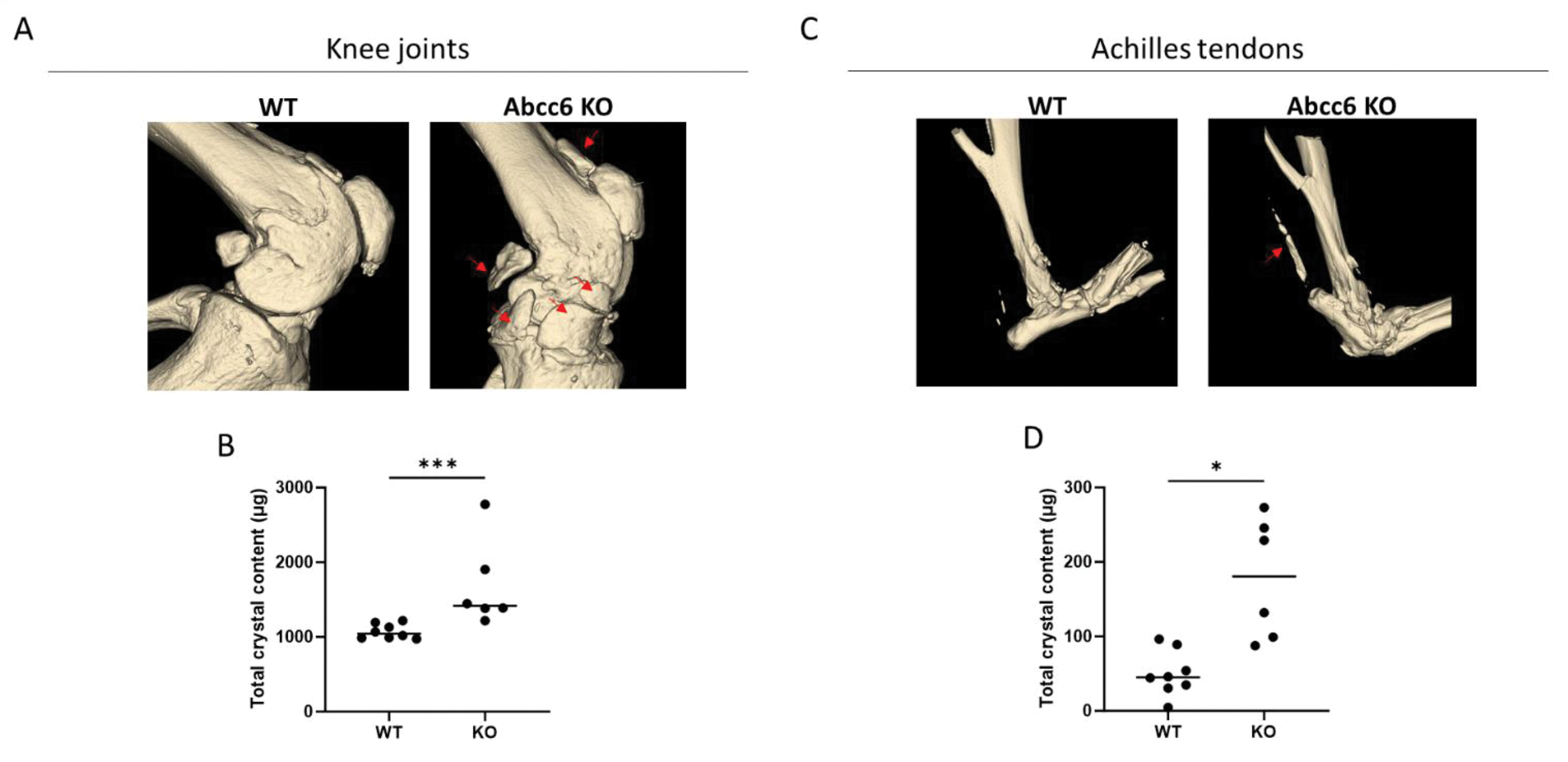
Results: In knee joints, calcification began at 16 weeks (mean crystal content of 778 µg in WT joints vs. 841 µg in KO joints, p=0.0284) and progressed to 43 weeks (mean crystal content of 1073 µg in WT joints vs. 1686 µg in KO joints, p=0.0007), as determined by micro-CT analysis. In 16-week-old mice we found no difference in cartilage damage and synovial inflammation. By contrast, in 43-week-old ABCC6-/- mice, increased knee calcification was accompanied by significantly greater cartilage degradation (mean score of 1.4 in WT vs. 4.3 in KO, p=0.01) and synovial inflammation (mean score of 0.2 in WT vs. 0.7 in KO, p=0.04). Calcification of the Achilles tendons was only evident in 43-week-old tissues, as seen in CT-scan images (mean crystal content of 50 µg in WT vs. 177.8 µg in KO, p=0.0101). We excluded that ABCC6 deficiency impacted bone mineralization as no significant changes were observed in tibial trabecular subchondral bone structure at 43 weeks (mean bone mineral density of 341.5 mg.cm 3 in WT vs. 349.8 mg.cm 3 in KO, mean trabecular thickness 0.016 cm vs 0.018 cm in KO, mean trabecular separation 0.018 cm vs 0.019 cm in KO, all non-significant).
Representative CT-scan images of 43-week-old WT and ABCC6 KO mice. (A-B) Knee joints; (C-D) Achilles tendons. Red arrows show increased calcifications in KO mice. *p=0.0101; ***p=0.0007.
Conclusion: Spontaneous and progressive calcification was observed in both knee joints and tendons of ABCC6-/- mice. This genetic model might provide a valuable tool for investigating disease mechanisms and identifying novel therapeutic strategies for OA and CT, for which current treatments remain ineffective.
REFERENCES: NIL.
Acknowledgements: NIL.
Disclosure of Interests: None declared.
© The Authors 2025. This abstract is an open access article published in Annals of Rheumatic Diseases under the CC BY-NC-ND license (