fetching data ... 
Background: Advances in Artificial Intelligence (AI) have opened new opportunities for improving detection, as well as the accuracy and efficiency of imaging interpretation in axial spondyloarthritis (axSpA). Timely and accurate diagnosis is essential for effective management of axSpA.
Objectives: To conduct a systematic literature review evaluating the performance of AI techniques in analyzing and interpreting imaging modalities (Magnetic resonance imaging (MRI), computed tomography (CT) and conventional radiography (CR)) for the assessment of axSpA.
Methods: In line with PRISMA guidelines, a comprehensive literature search was conducted across PubMed and Scopus for studies published between 1 January 2010 and 15 November 2024, using a search strategy adhering to pre-specified inclusion and exclusion criteria. First, two authors independently screened references based on titles and abstracts, followed by full-text review of the selected manuscripts. Disagreement was resolved by a third author. Study characteristics, AI techniques, and performance metrics (area under the receiving operating curve (AUC), sensitivity, specificity and accuracy) versus expert/human readers (reference standard), were extracted and summarized from the selected studies.
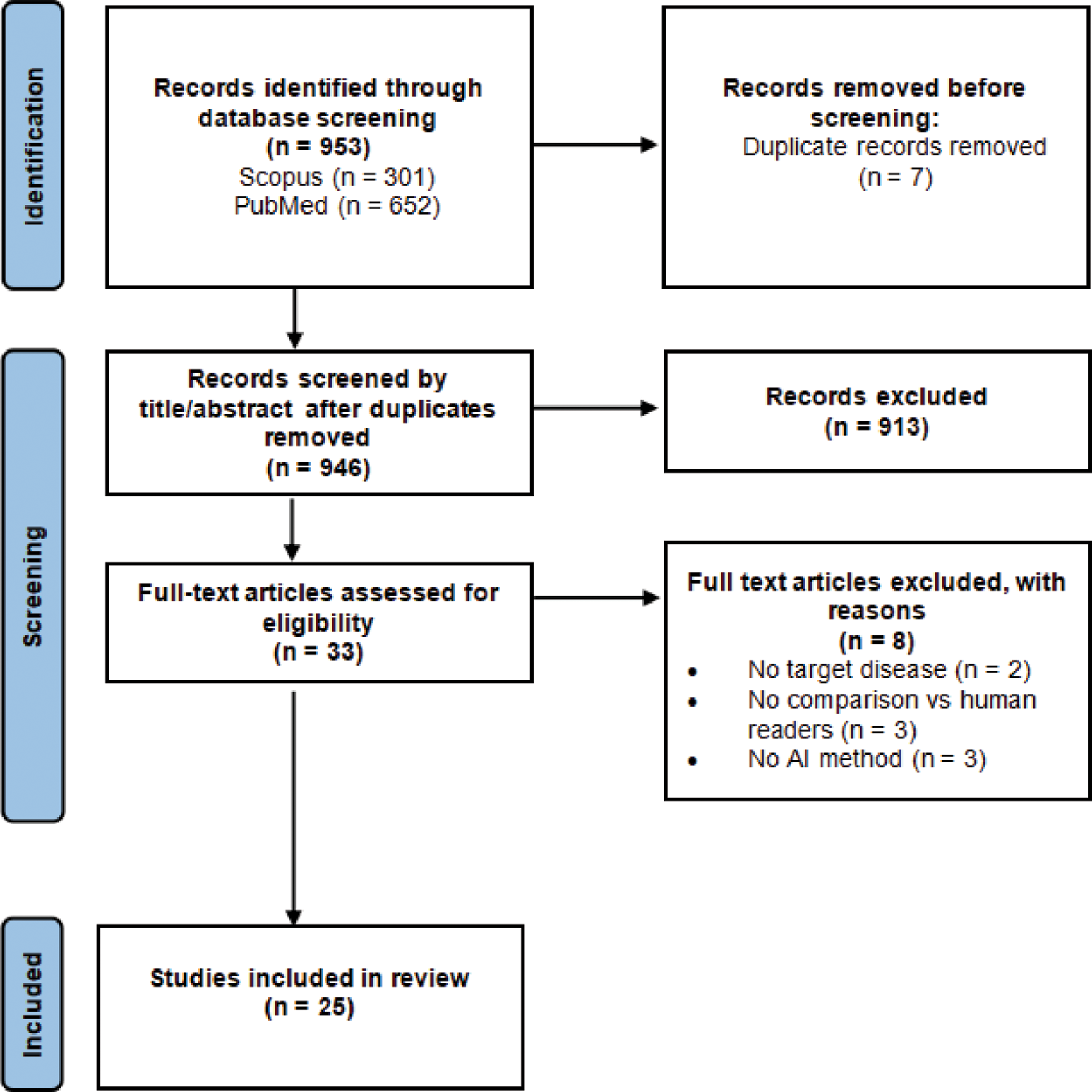
Results: A total of 953 references were identified, 33 full texts were reviewed, and 25 studies were included (PRISMA flowchart, Figure 1). All studies were published between 2020 and 2024, with 40% in 2024. Geographical distribution was as follows: Asia (15 studies), Europe (8 studies), Europe/North America (1 study) and South America (1 study). The number of subjects ranged from 56 to 2,011. Imaging methods included MRI (16 studies), radiography (6 studies), CT (2 studies) and MRI/radiography (1 study). AI performance was assessed using AUC in 13 studies, ranging from 74% to 97% (≥80% in 11 studies). Sensitivity, evaluated in 15 studies, ranged from 56% to 100% (≥80% in 12 studies, ≥90% in 7 studies). Specificity, assessed in 12 studies, ranged from 67% to 100% (≥80% in 10 studies, ≥90% in 5 studies). Accuracy, measured in 8 studies, ranged from 65% to 93% (≥80% in 7 studies, ≥90% in 3 studies). Individual results and overall estimates will be presented in Forest plots using meta-analyses to provide a quantitative summary measure on the performance metrics.
PRISMA Flowchart.
Conclusion: The number of studies assessing AI performance in axSpA using imaging modalities has increased tremendously in 2024, with over 50% of studies originating from Asia. MRI was the most frequently used imaging modality. In the majority of studies, AI performance was good (sensitivity/specificity ≥80%) to excellent (≥90%). This highlights the growing potential of AI to complement expert readers and improve the accuracy and efficiency of axSpA imaging assessment, particularly in MRI.
REFERENCES: NIL.
Acknowledgements: NIL.
Disclosure of Interests: Dominique Rosillon GSK shares, paid consultancy via DESiRE consulting, Manouk de Hooge paid consultancy for UCB Pharma, Gaëlle Varkas Speaker/consultancy fees: AbbVie, UCB, Pfizer, Eli Lilly, Novartis, Janssen, Galapagos, Celltrion. Educational grants: ASAS, KBVR-SRBR, Celltrion, EG, Grant research support: Takeda, Celltrion, Galapagos, AbbVie, MSD, Pfizer, Emilia Gvozdenovic Paid consultancy via DESiRE-consulting.
© The Authors 2025. This abstract is an open access article published in Annals of Rheumatic Diseases under the CC BY-NC-ND license (