fetching data ... 
Background: Promising systemic sclerosis (SSc) treatments may fail in clinical trials, in part due to inadequate measurement tools such as the modified Rodnan skin score (mRSS) that has variability between observers and does not fully assess the complex tissue microarchitecture abnormalities within SSc skin. A highly reproducible, quantitative tool to measure skin involvement in SSc is needed to improve clinical trial design and inform patient care decisions. Quantitative ultrasound (QUS) presents a unique opportunity to address this unmet need. Our novel, point-of-care (POC) high-frequency instrument was built to the specifications of SSc skin by analyzing >50 SSc skin biopsies and is equipped with two handheld probes operating at 50-MHz and 80-MHz. Unlike conventional US (1-10 MHz), and even other high-frequency US devices (30-20 MHz), the very high frequencies of our POC SSc-QUS instrument yields QUS parameters sensitive to tissue structures 6-10 µm in diameter, providing quantitative insight into the microstructure of the collagen fibrils in SSc skin. The device does not generate direct skin images, but rather numeric outputs that are reflective of key microarchitecture features relevant to SSc including edema, fibrosis, and collagen microstructural changes. Therefore, we hypothesize that compared to other US approaches, our POC instrument will be more sensitive and specific to the microstructural changes occurring in SSc.
Objectives: The purpose of this pilot, in-vivo study is to test QUS parameters as a tool to distinguish between SSc and healthy skin using the novel POC SSc-QUS device.
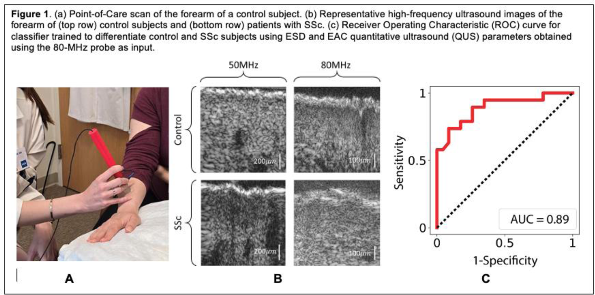
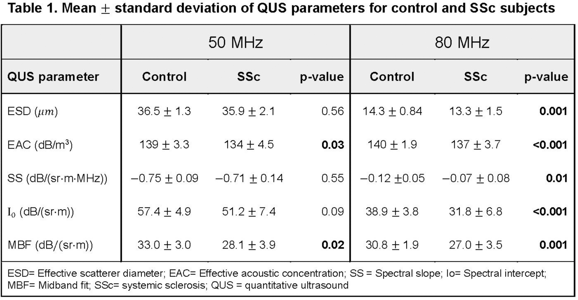
Methods: Healthy controls without inflammatory skin disease (N=8) and individuals with diffuse SSc (N=6) were recruited for POC-based high-frequency QUS (50, 80MHz) assessments of both forearms. Five backscatter coefficient-based QUS parameters, effective scatterer diameter (ESD), effective acoustic concentration (EAC), spectral slope (SS), spectral intercept (I0), and midband fit (MBF), that relate to tissue microstructure, were compared between SSc patients and controls using an independent-samples t-test. A support vector machine (SVM) classifier was trained using a radial basis function kernel and ESD and EAC as input features. Classifier performance was evaluated using a receiver-operating characteristic curve (ROC) and calculating the area under the ROC (AUC).
Results: Median (IQR) age of controls and SSc patients were 30 (25, 33) and 54 (48, 59), respectively. Median (IQR) total modified Rodnan Skin score (mRSS) was 24 (17, 34), local (US-site) mRSS was 2 (1, 3), and disease duration was 3.2 (1.4, 5.3) years. Representative high-frequency ultrasound images from control and SSc forearm skin are shown in Figure 1b. Significant differences in all five QUS parameters were noted between controls and SSc patients at 80 MHz (very high frequency), but only in EAC and MBF at 50 MHz (Table 1). A machine learning classifier trained using ESD and EAC computed from data acquired using the 80-MHz probe as inputs was able to differentiate SSc and control skin with a sensitivity, specificity, and AUC of 90%, 74%, 0.89, respectively (Figure 1c).
Conclusion: A highly accurate model using high-frequency (80MHz) QUS parameters was developed for classifying SSc versus control skin, indicating that 80MHz frequency is sensitive to detecting SSc-related microstructural changes. Studies are underway to correlate QUS parameters with SSc skin severity and progression in a larger patient cohort.
REFERENCES: NIL.
Acknowledgements: This work was supported by the Hospital for Special Surgery Discovery Award.
Disclosure of Interests: Kimberly Lakin: None declared, Cameron Hoerig: None declared, Jessica Gordon Merck, Cumberland Pharmaceuticals, Elmira Ghahramani: None declared, Shangke Liu: None declared, Mia Diaz: None declared, Robert Spiera AbbVie/Abbott, BMS, Regeneron, Amgen, Vera, GlaxoSmithKline, Novartis, Boehringer Ingelheim, Galderma, Sanofi, Roche Genentech, Cytori, AstraZeneca, Chemocentryx, Certa, AbbVie/Abbott, AstraZeneca, Chemocentryx, Corbus, Genetech, GlaxoSmithKline, InflaRx, Kadmon, Novartis, Principia, Roche, Sanofi, Cytori, Boehringer Ingelheim, Horizon Pharma, Dana Orange: None declared, Jonathan Mamou: None declared.
© The Authors 2025. This abstract is an open access article published in Annals of Rheumatic Diseases under the CC BY-NC-ND license (