fetching data ... 
Background: MM-II is a dispersion of empty large multilamellar liposomes for intraarticular (IA) injection and was recently shown to reduce knee pain in osteoarthritic patients for up to 26 weeks in a large global clinical study (NCT04506463). Previous in vivo studies demonstrated that these liposomes coat articular cartilage and menisci under compression load, provide boundary lubrication and reduce cartilage wear. Following IA injection, the liposomes are largely absorbed into the joint tissues where their lipids are metabolized and cleared from the joint cavity, while some are deposited on surfaces of knee components where they carry out their lubricative function.
Objectives: The aim of the current study was to characterize both the clearance profile of these liposomes and their association with various knee joint components following IA injection in healthy rats.
Methods: Sulfo-Cyanin 5.5-labelled MM-II (Cy5.5-MM-II) liposomes were prepared from dipalmitoylphosphatidylcholine (DPPC), dimyristoylphosphatidylcholine (DMPC) and Sulfo-Cy5.5- phosphatidylethanolamine at a molar ratio of 0.5445:0.4550:0.0005, respectively. Male Wistar rats (N=8) were bilaterally IA injected on day 0 with Cy5.5-MM-II (3.2mg per knee), scanned using a live imager (IVIS Lumina LT) on days 0, 1, 4, 6, 14 and 28 and the total radiant efficiency was determined in both knees. An additional non-injected rat served as a control of background autofluorescence. On day 28, the rats were euthanized and their knees isolated. To assess cartilage retention using IVIS, the knee joints from Cy5.5-MM-II-injected rats (N=3) and a control rat were cleaned of surrounding soft tissue. The knee joints were then disassembled, and femur head, tibia plateau, patella and menisci were isolated and scanned. Selection of regions of interest (ROIs) in cartilage areas of the femur, tibia and patella was done while being blinded to fluorescence signal to avoid bias. The signals were normalized to ROI area (average radiant efficiency). Corresponding signals in control rats were subtracted. The reported data are net signals normalized to that found in femurs at each time point. In addition, knees from MM-II -treated and control rats (n=2 knees each) were harvested, decalcified, stained with 4’,6-diamidino-2-phenylindole (DAPI) to visualize cell nuclei and used to prepare cryosections followed by fluorescent microscopy analysis.
Results: MM-II clearance from knee joints displayed a biphasic profile: an initial first-order exponential phase during the first 11 days followed by a terminal phase reaching a quasi-state to a value of with a value of about 3% of injected dose which remained at this value through the end of the study (Figure 1A). MM-II half-life in the exponential phase was about 3 days. On day 28, ex vivo analysis was performed on intact knees and on isolated femurs, tibias and patellae from MM-II injected rats as well as from the non-injected control rat. Representative images of knees and knee parts from MM-II-injected and control rats showed MM-II-associated signal in both intact knees, as well as in all isolated knee parts (Figure 1B). Analysis of the signals relative to that found in the femur cartilage showed that these liposomes distribute in cartilage of femur, tibia and patella to a similar extent (Figure 1C). Notably, in addition to their retention in cartilage, MM-II liposomes were also found in menisci (Figure 1 B and 1C). Histological analysis supported the ex-vivo analysis and showed that MM-II coating of cartilage and menisci persisted at least for 28 days post injection (end of the follow-up period).
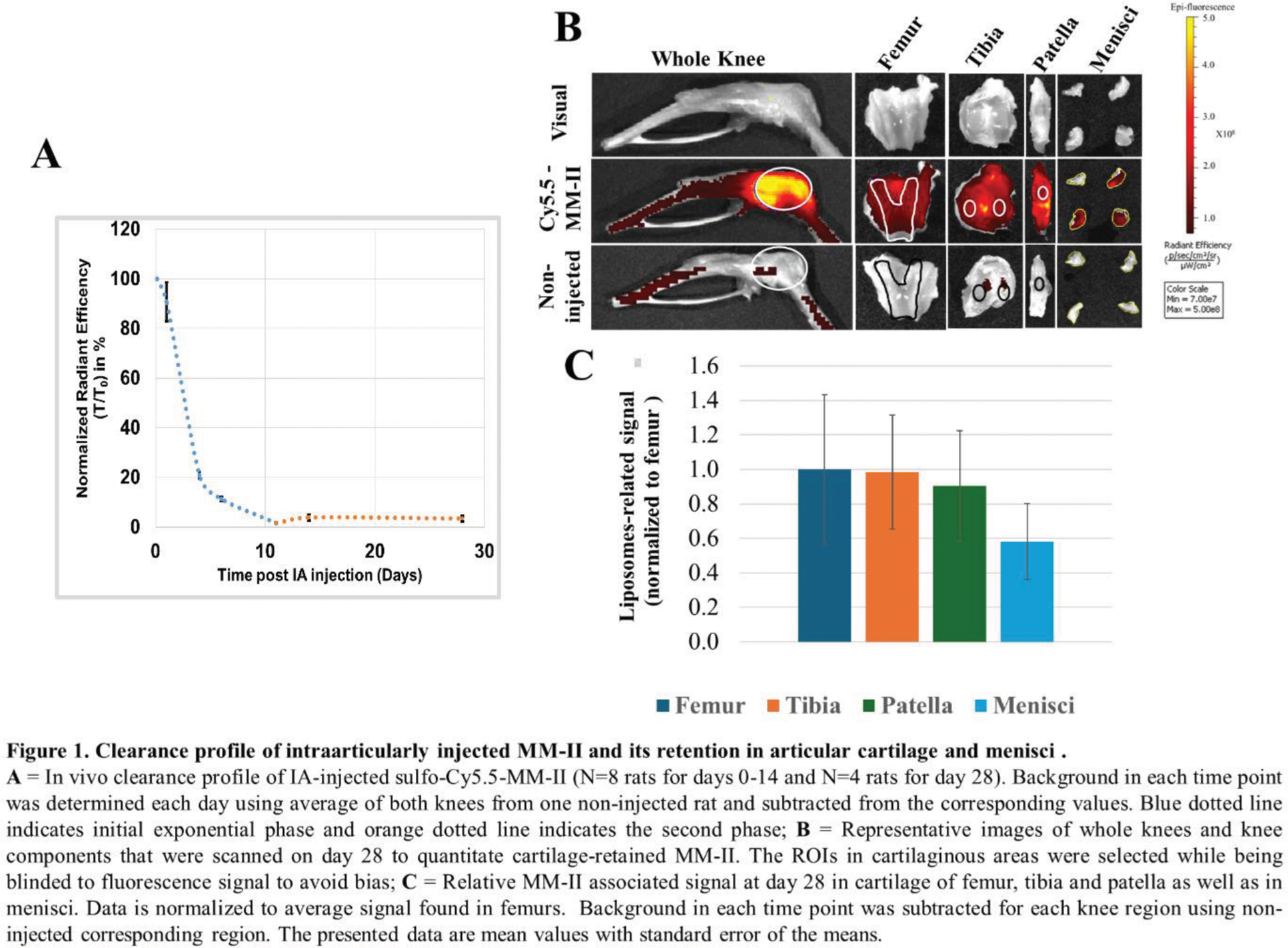
Conclusion: Following IA injection into healthy rat knees, a significant part of the injected MM-II liposomes cleared from knee joints within 1-2 weeks post-injection, likely due to uptake by synovial macrophages. Yet, a persistent amount of the liposomes remained associated with articular cartilage and menisci for at least 28 days. This data provides mechanistic support for the recent finding in a phase2b clinical trial showing reduction of knee pain lasting up to 6 months.
REFERENCES: NIL.
Acknowledgements: NIL.
Disclosure of Interests: Roni Wechsler I received options of Moebius Medical as an employee, Current employee of Moebius Medical, Before joining Moebius Medical, I served as a consultant for Moebius Medical, Rany Rotem Current employee of Moebius Medical.
© The Authors 2025. This abstract is an open access article published in Annals of Rheumatic Diseases under the CC BY-NC-ND license (