fetching data ... 
Background: CCN1, a matricellular protein with angiogenic and immunomodulatory properties, is overexpressed in endothelial and synovial tissues of rheumatoid arthritis (RA) patients. Previous findings demonstrated that CCN1 inhibition reduces synovial angiogenesis and structural damages in preclinical arthritis models. However, it remains unclear whether these therapeutic effects of CCN1 inhibition are mediated through a reduction in angiogenesis.
Objectives: This study investigates the ability of Cilengitide, a cyclic pentapeptide integrin inhibitor, to block CCN1-mediated effects on endothelial cells (ECs) and evaluates its potential therapeutic impact in the TNF-dependent Tg197 arthritis model.
Methods: The effects of CCN1 inhibition on angiogenesis through Cilengitide were assessed in vitro using ECs derived from circulating progenitors. Cells were cultured on Matrigel with CCN1 (0.2 µg/ml), either alone or with increasing doses of Cilengitide (2.5–10 µM), and capillary-like tube formation was analyzed. For in vivo studies, TNF-transgenic mice (Tg197) received intraperitoneal injections of either Cilengitide (10 mg/kg) or PBS three times weekly. Clinical arthritis scores were monitored bi-weekly, and after six weeks, paws were analyzed using micro-CT and histology. Immune profiling from the spleen of TG197 mice was performed via flow cytometry.
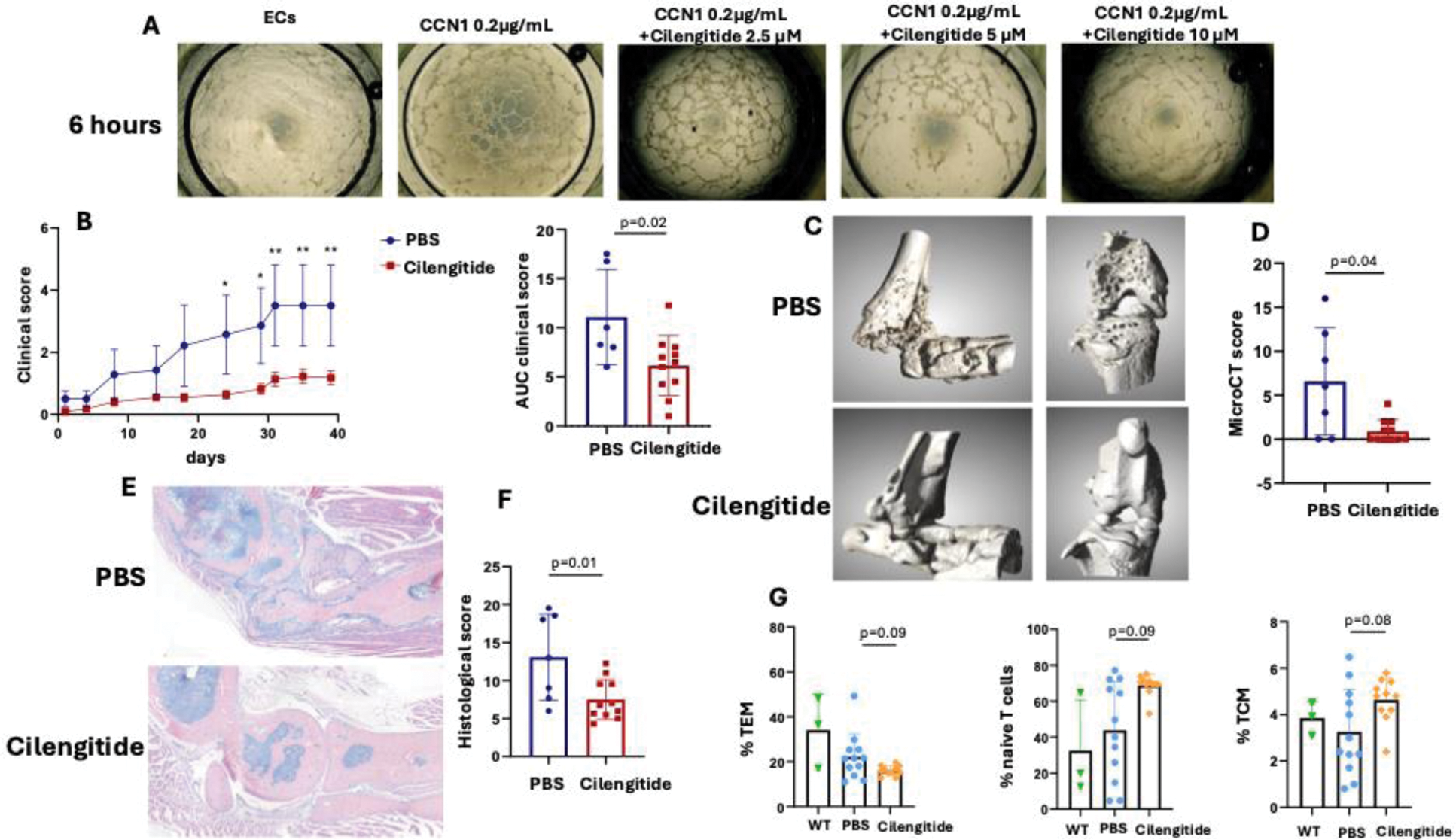
Results: In vitro , CCN1 induced robust capillary-like tube formation, which was significantly inhibited by Cilengitide in a dose-dependent manner. At 2.5 µM, tube formation was reduced, and at 5–10 µM, nearly complete inhibition was observed (Figure 1A), strongly supporting that CCN1 effects on ECs are mediated through integrins αvβ3 and αvβ5, which are critical for angiogenesis. In vivo , Cilengitide significantly improved clinical arthritis scores (Figure 1B). The AUC of the clinical score of Cilengitide-injected mice decreased significantly by 55±6% (p=0.020). Micro-CT analysis demonstrated better-preserved joint integrity in Cilengitide-treated mice, with markedly fewer bone erosions compared to controls (Figure 1C-D). Histological analysis of hind paw sections confirmed previous results, with significantly less inflammation and structural damage in Cilengitide-treated mice (Figure 1E-F). Flow cytometry showed decreased activation of CD4+ T cells in Cilengitide treated mice, with reduced PD-1 expression, indicating lower exhaustion levels. CD4+ T cells exhibited a shift in memory subsets following Cilengitide treatment, with fewer effector memory T cells and an increase in central memory and naïve T cells (Figure 1G).
Conclusion: Cilengitide effectively inhibits CCN1-induced angiogenesis in vitro , confirming that CCN1 exerts its effects on endothelial cells through integrins αvβ3 and αvβ5. In vivo , Cilengitide significantly reduces clinical, structural, and histological damage in the Tg197 arthritis model while modulating immune profiles. These findings suggest that CCN1’s effects in RA, including structural and inflammatory damage, are mediated by its role in promoting pathological angiogenesis. Targeting CCN1 and associated pathways offers a promising therapeutic strategy for RA, complementing current treatments.
Effects of CCN1 inhibition by Cilengitide on tube formation and experimental arthritis
A, Representative images of tube formation at 6 hours in RA endothelial cells (ECs) alone, or following treatment with recombinant CCN1 (0.2 µg/mL) with or without Cilengitide (2.5, 5 or 10 µM) (4x magnification). B, Experimental arthritis in Tg197 mice receiving IP injections of Cilengitide (10 mg/kg) or PBS (100 µL) 3 times weekly. Y-axis shows clinical score and the area under the curve (AUC) of the clinical score (PBS: n=7 mice, Cilengitide, n=11 mice). C, Representative 3D reconstructions of ankles, tarsus and knees of Tg197 mice treated with Cilengitide or PBS. D, Y-axis shows the mean erosion score (from 0 to 16). E, Sections of ankle and tarsus joints from Tg197 mice receiving Cilengitide or PBS stained with hematoxylin (scale bar = 200µm). F, Histomorphometric analysis of the area of synovitis and bone destruction. G, Flow cytometry analysis of T cell subsets (effector memory T cells, TEM, naive T cells and central memory T cells, TCM) from the spleen of wild type (WT) mice (n=3) and Tg197 mice treated with Cilengitide or PBS. All data are shown as the means ± SD. Statistical tests: Student’s t test for 2-group comparisons, ANOVA with Dunnett’s multiple comparisons test for 3-group comparisons. Data are representative of a single experiment.
REFERENCES: NIL.
Acknowledgements: NIL.
Disclosure of Interests: None declared.
© The Authors 2025. This abstract is an open access article published in Annals of Rheumatic Diseases under the CC BY-NC-ND license (