fetching data ... 
Background: Aging affects all physiological functions of the human body, including the immune system, and most types of cells in the innate immune system and the adaptive immune system may undergo age-related changes. Rheumatoid Arthritis (RA) is a highly disabling disease that occurs most frequently in elderly patients. Recently, a type of CD4+T cell with cytotoxic phenotype and B-cell auxiliary function has been found to accumulate with Age and is associated with the activity of systemic lupus erythematosus, which is defined as age-associated helper T cells (ThA). However, the correlation between ThA cells and RA and the possible key regulatory factors have not been identified.
Objectives: This study aims to understand the distribution of ThA cells in RA patients and explore potential key signaling pathways and intercellular communication patterns, which may provide new evidence to explain the pathogenesis and progression of rheumatoid arthritis.
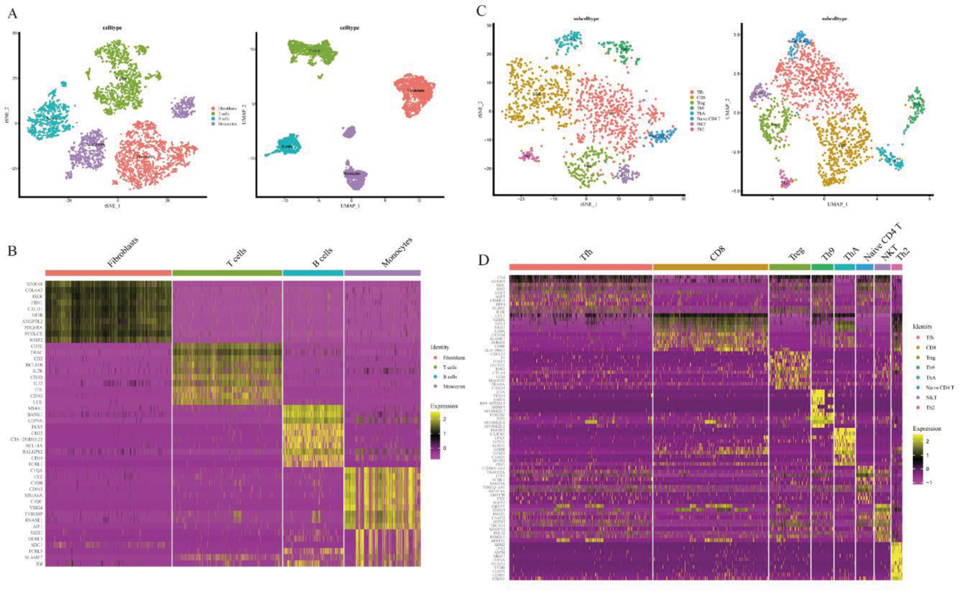
Methods: We used 5172 cells from the published synovial tissue single-cell sequencing dataset SDY998, of which 4525 cells were measured in patients with RA and 647 cells in patients with OA. Then, Seurat and CellChat software packages in R 4.1.3 were used to conduct quality control, dimensionality reduction, clustering, grouping, and cell annotation on the single cell transcriptome dataset, and T cells were re-grouped to further analyze the intercellular communication mode.
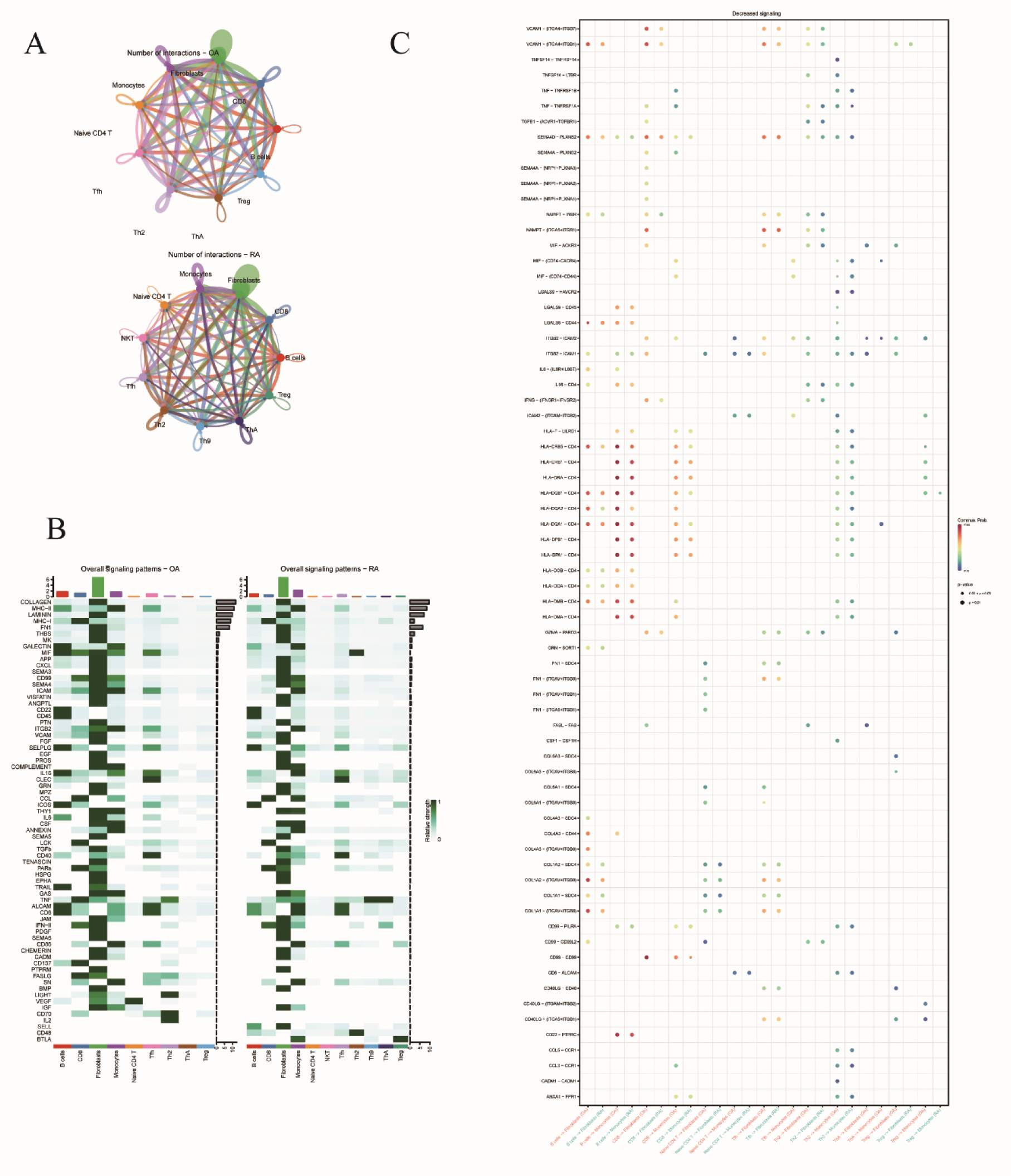
Results: Based on 5,172 cells, four cell types were initially identified: T cells (1,523), B cells (837), fibroblasts (1,754) and macrophages (1,058). T cells were further divided into 8 subsets of Tfh (564), CD8+ T (453), Treg (163), Th9 (87), ThA (81), Naive CD4+ T (69), NKT (63), Th2 (43). Signal pathway analysis showed that MHC-I, TNF, IFN-II, SN and other signaling pathways were highly expressed in RA patients. In RA patients, compared with OA patients, the number of cell communication increased, but the intensity decreased. When ThA was used as the ligand and fibroblasts were used as the receptor, Intercellular communication showed VCAM1-(ITGA4+ITGB7), VCAM1-(ITGA4+ITGB1), TNF-TNFRSF1A, TGFβ1-(ACVR1+TGFβR1), SEMA4D-PLXNB2, NAMPT-INSR, IFNγ-(IFNγR1+I) Fγr2), GZMA-RARD3 and COL1A2-SDC1 were highly expressed in RA patients.
Conclusion: This study confirmed the increased number of ThA cells in RA patients and the existence of cell-to-cell interactions with fibroblasts, suggesting that ThA cells are associated with RA disease. At the same time, it provides cell communication types and key signaling pathways between ThA cells and fibroblasts in RA patients, providing a research basis for studying the diagnosis and treatment of ThA in RA patients, but the specific mechanism of action still needs further research.
Dimension reduction, clustering and group identification of synovial tissue cell population and synovial tissue T cell population.
Analysis of communication between ThA cells and fibroblasts in rheumatoid arthritis (RA) and osteoarthritis (OA).
REFERENCES: [1] Tanemoto S, Sujino T, Miyamoto K, Moody J, Yoshimatsu Y, Ando Y, Koya I, Harada Y, Tojo AO, Ono K, Hayashi Y, Takabayashi K, Okabayashi K, Teratani T, Mikami Y, Nakamoto N, Hosoe N, Ogata H, Hon CC, Shin JW, Kanai T. Single-cell transcriptomics of human gut T cells identifies cytotoxic CD4+CD8A+ T cells related to mouse CD4 cytotoxic T cells. Front Immunol. 2022 Oct 24;13:977117.
[2] Binvignat M, Miao BY, Wibrand C, Yang MM, Rychkov D, Flynn E, Nititham J, Tamaki W, Khan U, Carvidi A, Krueger M, Niemi E, Sun Y, Fragiadakis GK, Sellam J, Mariotti-Ferrandiz E, Klatzmann D, Gross AJ, Ye CJ, Butte AJ, Criswell LA, Nakamura MC, Sirota M. Single-cell RNA-Seq analysis reveals cell subsets and gene signatures associated with rheumatoid arthritis disease activity. JCI Insight. 2024 Jul 2;9(16):e178499.
Acknowledgements: Thanks to my family and my cats.
Disclosure of Interests: None declared.
© The Authors 2025. This abstract is an open access article published in Annals of Rheumatic Diseases under the CC BY-NC-ND license (