fetching data ... 
Background: Idiopathic inflammatory myopathies (IIM) are characterized especially by inflammatory involvement of muscles. MicroRNAs (miRNAs), such as miR-133a-3p and miR-1-3p, are small, non-coding RNA molecules that regulate gene expression by binding to target mRNAs, leading to mRNA degradation or inhibition of protein translation, and play an important role in regulating gene expression, as well as muscle tissue repair. Their dysregulated expression has been implicated in IIM pathogenesis, highlighting their potential as biomarkers and therapeutic targets.
Objectives: To assess the changes in the expression of miR-133a-3p and miR-1-3p in IIM, and their association with muscle involvement, disease-related features, and the effect of initial immunosuppressive therapy.
Methods: 66 patients with IIM were included: 44 females; mean (SD) age 57.6 (13.9) years; disease duration 2.3 (3.0) years; 21 dermatomyositis (DM)/29 polymyositis (PM), both without statin history/16 immune-mediated necrotizing myositis statin-induced (IMNM). 15 IIM (6 DM, 6 PM, 3 IMNM) had a follow-up 6 months after immunosuppressive therapy initiation to analyze the impact of treatment on miRNA expression. In all patients, basal laboratory parameters, disease activity, damage, and muscle involvement were evaluated (Myositis Intention to Treat Index (MITAX), Myositis Damage Index (MDI), Manual Muscle Testing-8 (MMT-8)), comorbidities, and current treatment were recorded. Body composition was examined using bioelectric impedance analysis (BIA-2000-M). Total RNA was extracted from plasma using the miRNeasy Serum/Plasma Advanced kit (Qiagen). Quantitative PCR was performed using 6 miRCURY LNA miRNA PCR assays (Qiagen) with miR-103-3p, miR-191-5p, and let-7a-5p as endogenous controls. miRNA concentration analysis was performed using GenEx software (Multid Analysis AB). Data are presented as mean (SEM).
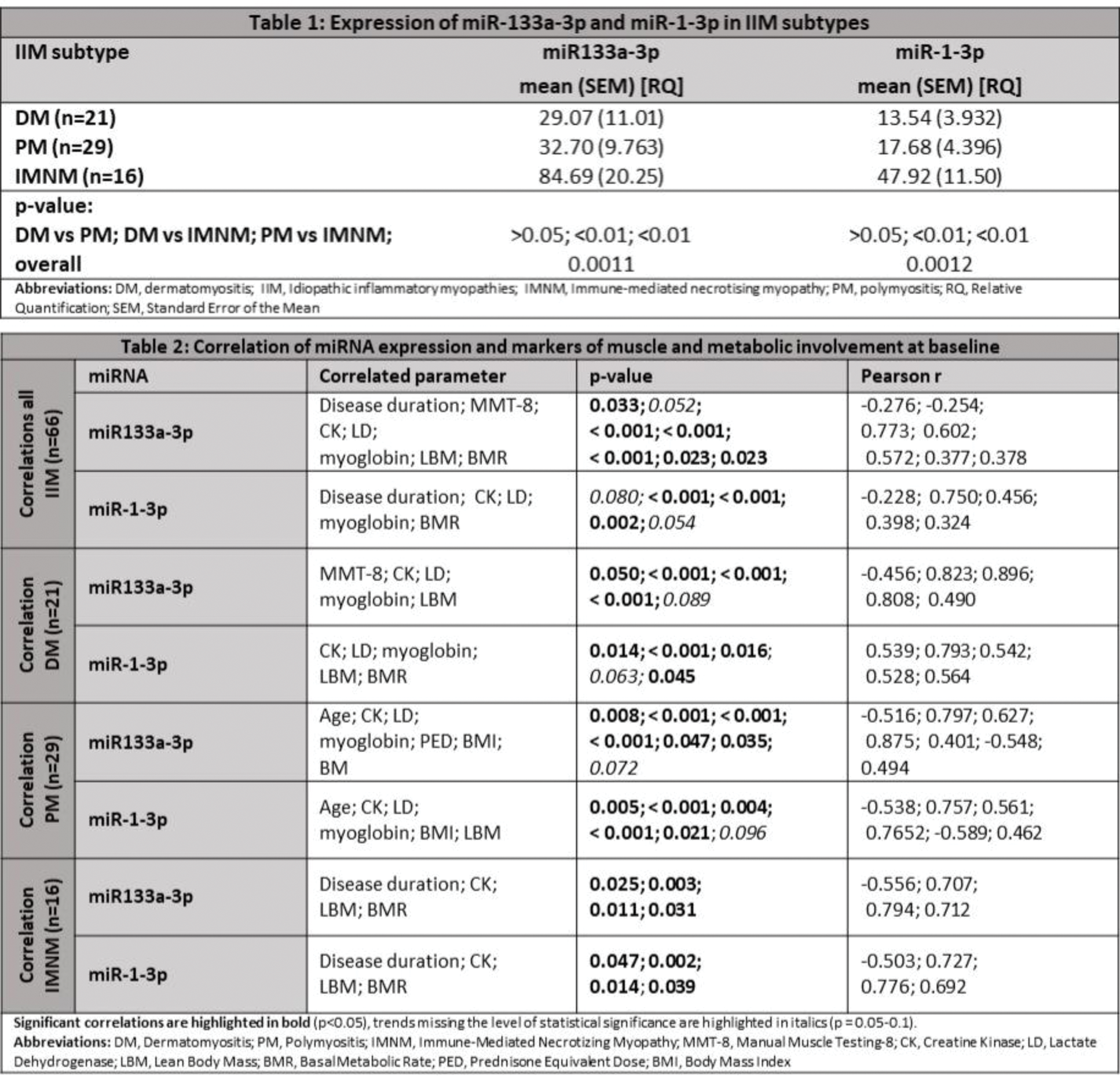
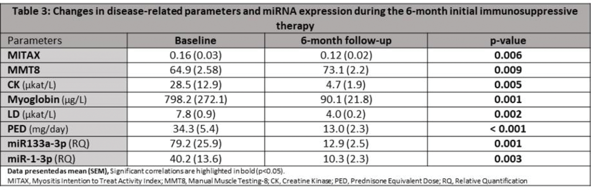
Results: The expression of miR-133a-3p and miR-1-3p was significantly higher in IMNM patients compared to DM and PM (Table 1). Elevated levels of miR-133a-3p and miR-1-3p were associated with increased markers of muscle damage (CK, LD, and myoglobin). In addition, increased levels of miR-133a-3p were also associated with shorter disease duration, higher amount of muscle tissue (lean body mass) and higher basal metabolic rate (both by BIA) and borderline with decreased muscle strength (Table 2). A significant decrease in expression of miR-133a-3p and miR-1-3p was observed 6 months after the initiation of immunosuppressive therapy, which caused a significant decrease in the disease activity (MITAX) and the serum markers of muscle damage and an increase in muscle strength (MMT-8) (Table 3). A larger decrease in the expression of miR-133a-3p was associated with a larger decrease in CK (p<0.0001; r=0.901) and LD (p=0.012; r= 0.696) and borderline with a larger improvement in muscle strength (p=0.052; r=0.572). Similarly, a more pronounced decrease in the expression of miR-1-3p was associated with a marked decrease in CK (p=0.0004; r=0.852) and borderline with larger muscle strength (p=0.071; r=0.538) and decrease in LD (p=0.105; r=0.491). Higher baseline levels of miR-133a-3p and miR-1-3p predicted greater improvements in muscle strength (p=0.048; r=0.578 and p=0.048; r=0.580) and a larger decrease in CK (p=0.0002; r=0.878 and p=0.0012; r=0.816) and LD levels (p=0.009; r=0.709 and p=0.091; r=0.509).
Conclusion: The expression of both miR-133a-3p and miR-1-3p was shown to be significantly higher in IMNM patients, compared to DM and PM, indicating more pronounced muscle damage in IMNM. Expression of both miRNAs appeared to be most increased in the early disease characterized with high disease activity and muscle damage and declined in 6 months upon the initial immunosuppressive therapy in line with the decline in serum markers of muscle damage and with the improvement of muscle strength.Our study demonstrated both miR-133a-3p and miR-1-3p as a potential biomarker of disease activity and muscle damage in IIM, and a potential predictor of initial treatment response in IIM.
REFERENCES: NIL.
Acknowledgements: Supported by MH CR 023728, NU21-01-00146, NU21-05-00322, Ministry of Education Youth and Sports of the Czech Republic [BBMRI.cz-LM2023033], and SVV 260638.
Disclosure of Interests: None declared.
© The Authors 2025. This abstract is an open access article published in Annals of Rheumatic Diseases under the CC BY-NC-ND license (