fetching data ... 
Background: Melanoma differentiation–associated gene 5 (MDA5) is a member of the RIG-I–like receptor family and functions as a cytosolic sensor of viral double-stranded RNA. Anti–MDA5 antibody–positive dermatomyositis (DM) has recently been shown to be strongly associated with lethal rapidly progressive interstitial lung disease (RP-ILD). In addition, approximately 20% of patients develop early liver dysfunction. We previously demonstrated deposition of immunoglobulins and complement component C3 in lung tissues from patients with anti–MDA5 antibody–positive DM–associated ILD [1], suggesting a pathogenic role for antibody- and complement-mediated tissue injury in this disease. Furthermore, anti–MDA5 antibody–positive DM cases have been reported to exhibit increased serum levels of soluble CD206 and enhanced infiltration of CD206-positive macrophages in lung tissue. These findings suggest that M2-like macrophages may play a pivotal role in mediating tissue injury and fibrosis in anti–MDA5 antibody–positive DM.
Objectives: This study aimed to investigate the mechanisms of hepatic injury and the role of macrophages and complement system in antibody-positive DM using human autopsy cases and a human MDA5 transgenic (Tg) mouse model.
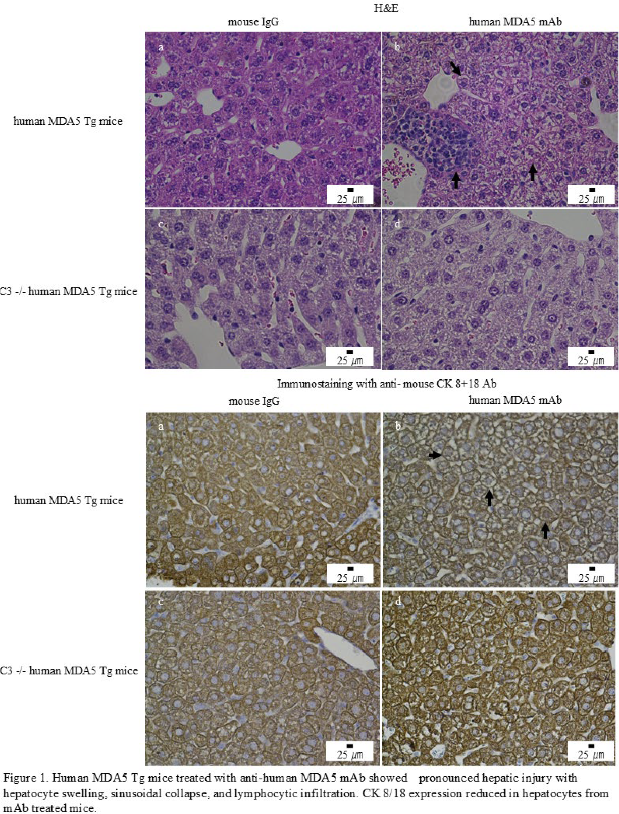
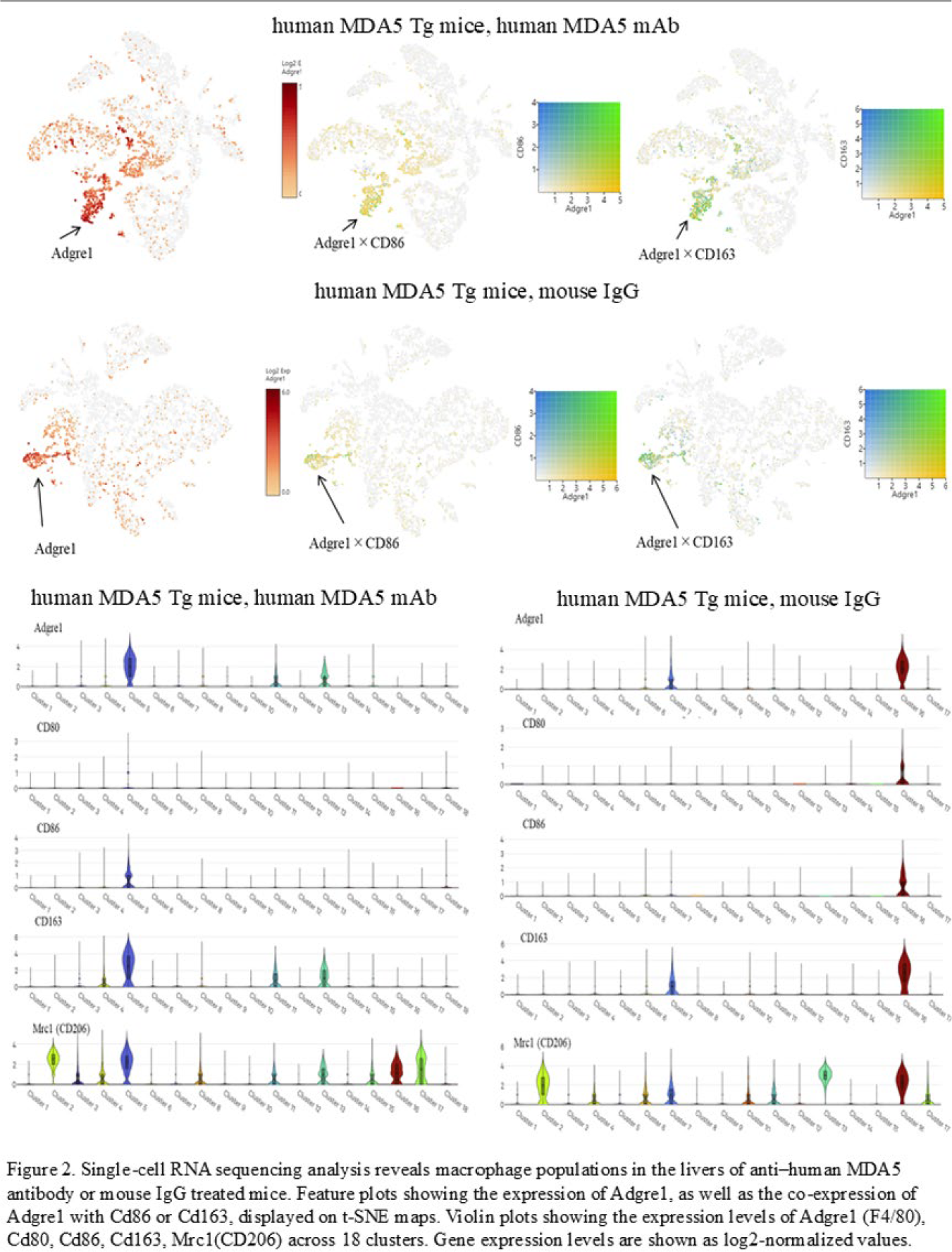
Methods: Liver specimens from five autopsy cases of anti–MDA5 antibody–positive DM were analyzed histopathologically. To establish a model of antibody-mediated hepatic injury, human MDA5 Tg mice were treated with anti-human MDA5 monoclonal antibodies (mAbs). The contribution of complement was assessed by comparing hepatic pathology between wild-type C3 (+/+) and C3-deficient (−/−) MDA5 Tg mice. Liver tissues were analyzed by immunohistochemistry, western blotting, and single-cell RNA sequencing. Human MDA5 Tg mice were treated with 0.5 mg of anti-human MDA5 mAb or control mouse IgG on days 0, 7, 14, and 21 and were sacrificed on day 28 for histological analysis and single-cell RNA sequencing. Dissected organs were subjected to immunohistochemical staining with anti-F4/80 mAb, anti-CD80 mAb, anti-CD206 polyclonal antibody, and anti–cytokeratin (CK) 8/18 antibody. Western blot analysis was performed using liver tissues from C3 (+/+) or C3 (−/−) human MDA5 Tg mice treated with anti–human MDA5 mAb or control mouse IgG. Protein expression levels were normalized to β-actin. Single-cell RNA-sequencing library preparation from frozen mouse livers of human MDA5 Tg mice treated with anti–human MDA5 mAb or control mouse IgG. For single-cell RNA sequencing, approximately 25 mg of liver tissue obtained from three individual mice in the anti-human MDA5 mAb-treated group and three mice in the control IgG-treated group were dissociated and pooled separately prior to library preparation, generating one mixed sample per group. Major cell clusters were annotated based on canonical marker genes followed by previously reported [2]. Macrophage-lineage cells annotated by Adgre1 (F4/80) cells, co-expressing Adgre1 (F4/80) and Cd86 or Adgre1 (F4/80) and Cd163 were operationally classified as M1-like and M2-like macrophages.
Results: In five anti–MDA5 antibody–positive DM autopsy cases, inflammatory cell infiltration was observed, whereas hepatic architecture and sinusoidal structures were preserved. Immunostaining for CK8/18 showed preserved expression in hepatocytes. F4/80-positive, CD80-positive, and CD206-positive macrophage-like cells were detected. Human MDA5 Tg mice treated with anti–human MDA5 mAb developed pronounced hepatic injury characterized by hepatocyte swelling, sinusoidal collapse, and lymphocytic infiltration. CK8/18 expression was preserved in control IgG–treated mice but reduced in mAb-treated mice. In contrast, hepatic injury was markedly attenuated in C3 (−/−) human MDA5 Tg mice treated with anti–human MDA5 mAb, with preserved CK8/18 expression. These findings indicate that complement activation contributes to anti–MDA5 antibody–induced hepatic injury (Figure 1). Immunohistochemistry demonstrated increased infiltration of F4/80-positive and CD206-positive M2-like macrophages in the livers of mAb-treated human MDA5 Tg mice. Western blot analysis revealed significantly higher hepatic F4/80 protein levels (p < 0.05) in anti–human MDA5 mAb–treated C3 (+/+) mice compared with control IgG–treated mice and C3 (−/−) mice treated with anti–human MDA5 mAb. Single-cell RNA sequencing identified a distinct macrophage cluster expressing Adgre1 in mAb-treated mice. Violin plot analysis demonstrated increased expression of M2-associated markers Cd163 and Mrc1, together with expression of M1-associated markers Cd80 and Cd86. In contrast, macrophages from control IgG–treated mice showed low expression of these markers (Figure 2).
Conclusions: Complement activation and the accumulation of M2-like macrophages are associated with anti-human MDA5 mAb–induced hepatic injury in mice. These findings provide mechanistic insight into antibody–complement–macrophage interactions and suggest that modulation of complement pathways may represent a potential therapeutic approach to limit liver and systemic involvement in this disorder.
REFERENCES: [1] Zaizen Y. et al. Enhanced immune complex formation in the lungs of patients with dermatomyositis. Respir Res (2023) 24(1): 86. doi: 10.1186/s12931-023-02362-0.
[2] Su Q. et al. Single-cell RNA transcriptome landscape of hepatocytes and non-parenchymal cells in healthy and NAFLD mouse liver. iScience (2021) 24(11): 103233. doi: 10.1016/j.isci.2021.103233.
Acknowledgments: NIL.
Disclosure of Interests: Takuma Koga: None declared, Yoshiaki Zaizen Argenx, Hiroyuki Suzuki: None declared, Suzuna Sugi: None declared, Hironao Hozumi: None declared, Noriho Sakamoto: None declared, Takafumi Suda: None declared, Hiroshi Mukae: None declared, Hironori Kusano: None declared, Akihiko Kawahara: None declared, Jun Akiba: None declared, Takumi Kawaguchi: None declared, Shinjiro Kaieda: None declared, Tomoaki Hoshino Argenx.