fetching data ... 
Background: Structural joint damage assessed on plain radiographs remains a key outcome in rheumatoid arthritis (RA) and psoriatic arthritis (PsA). Semi-quantitative scoring by expert readers of bone erosions (BE) and joint space narrowing (JSN) is the current regulatory standard but has limited sensitivity to change [1-3]. Quantitative measures may provide a more sensitive, reproducible assessment of structural progression.
Objectives: Building on the van der Heijde-modified Sharp (vdHS) framework, we developed and evaluated quantitative measures of BE area and joint space width (JSW) from hand radiographs using expert manual annotations to provide continuous measures of structural joint damage.
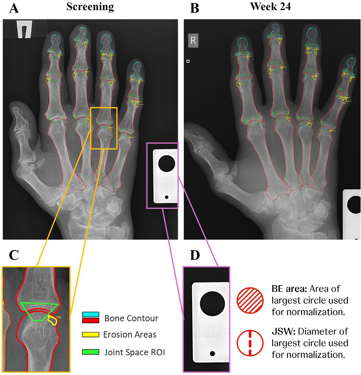
Methods: A pilot dataset of 100 paired right-hand radiographs (Screening and Week 24) from historical trial PsA patients selected to represent a range of vdHS severities was analyzed. These subjects were drawn from both placebo and active treatment arms. Manual annotations included delineation of 12 joints (distal interphalangeal joints 2–5, proximal interphalangeal joints 2–5, metacarpophalangeal joints 2-5) (Figure 1A–C). Initial contouring was first performed by a trained technician followed by a musculoskeletal radiologist experienced with vdHS scoring. Ground truth included joint level BE and JSN scores assigned by an expert panel using the vdHS scoring system modified for PsA. From these manual annotations, quantitative measures of joint structure were derived. For JSW, bone contour points were first filtered to retain only points within each predefined joint-space region of interest. The retained contours were smoothed with a fourth-degree polynomial and JSW across the joint was computed as the nearest-neighbor Euclidean distance between the bone surfaces. Six descriptive JSW metrics were calculated per joint: minimum, maximum, mean, 5th percentile, 10th percentile, and 15th percentile. For BE area, the summed area of manually delineated erosions within each joint was calculated. To account for varying image resolutions between visits and patients, all measures were normalized to the radiographic resolution marker (Figure 1D). Aggregate image-level scores were obtained by summing across all annotated joints. Finally, image-level BE and JSW scores were then compared with the expert-annotated aggregate BE and JSN scores within the same joints using Pearson correlations, with significance set at p<0.05. The distribution of within-subject change (Week 24 – Screening) for BE score and BE area as well as JSN score and JSW were visualized using empirical cumulative distribution functions. The proportion of subjects with worsening values per measure was determined.
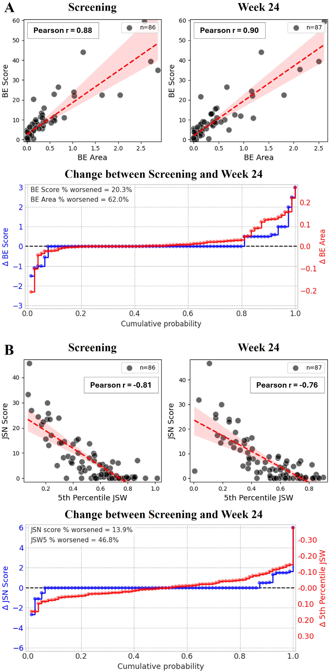
Results: After excluding images without a resolution marker, 86 radiographs at Screening and 87 radiographs at Week 24 were included in the final analysis. Image-level BE area correlated strongly with vdHS BE scores at both timepoints (Screening: r = 0.88, p < 0.001; Week 24: r = 0.90, p < 0.001; Figure 2A). Of the six JSW descriptors, the 5th percentile JSW showed the strongest correlation with vdHS JSN scores and is therefore presented as the representative measure. Image-level 5th percentile JSW measures showed strong negative correlations with vdHS JSN scores at both timepoints (Screening: r = -0.81, p < 0.001; Week 24: r = -0.76, p < 0.001; Figure 2B). Among the 79 patients with paired radiographs, BE score worsened in ~20% and BE area in 62% of patients (Figure 2A). JSN worsened in ~14% of patients (defined as an increase in JSN), while the 5th percentile JSW worsened in ~47% of patients (defined as a decrease in JSW) (Figure 2B).
Conclusions: This study demonstrates a quantitative framework for deriving continuous measures of BE area and JSW directly from plain radiographs that strongly correlate with image level vdHS scores. The continuous measures detected substantially more structural deterioration compared to the ordinal BE and JSN scores, suggesting they may identify earlier or subtler progression and hold promises for improving sensitivity in future trials aimed at measuring structural progression. However, interpretation should remain cautious given the modest paired sample size. Future work will assess clinical relevance and automate segmentation and landmark detection to enable scalable, observer-independent analysis.
Manual annotations and normalization for quantitative radiographic measures. Annotated structures include 16 bone contours (DP2-5, MP2-5, PP2-5, MC2-5), 12 joint-space ROIs (DIP2-5, PIP2-5, MCP2-5) and erosion contours. Panels show Screening (A) and Week 24 (B) radiographs and annotations from the same patient. (C) Zoomed view of a single joint. (D) Resolution marker and normalization used for BE area and JSW. A color legend is provided in the figure.
Relationship between quantitative image-level measures and vdHS scores. (A) Scatter plot of BE area versus vdHS BE score at Screening and Week 24 with fitted regression lines; accompanying empirical cumulative distribution functions (ECDFs) show within-subject changes for BE score and BE area. Data points represent individual images at each time point. (B) Scatter plot of 5th-percentile JSW versus vdHS JSN score; accompanying ECDFs show within-subject change in JSN score and 5th-percentile JSW (lower JSW indicates worsening; note inverted y-axis for JSW). Data points represent change observed in each patient. The dashed horizontal line represents no-change line and %-worsened annotations.
REFERENCES: [1] van der Heijde D. How to read radiographs according to the Sharp/van der Heijde method. J Rheumatol. 2000.
[2] van der Heijde D et al. Psoriatic arthritis imaging: a review of scoring methods. Ann Rheum Dis. 2005.
[3] Koc GH et al. Determinants of radiographic progression in early psoriatic arthritis: insights from a real-world cohort. RMD Open. 2024.
Acknowledgments: NIL.
Disclosure of Interests: Dimitri Kessler was an employee of J&J, Zijun Gao may own stock or stock options in J&J, employee of J&J, Nicholas Fountoulakis may own stock or stock options in J&J, employee of J&J;, Kathleen Wuyts may own stock or stock options in J&J, employee of J&J, Xiaoqin Tang may own stock or stock options in J&J, employee of J&J, Luiza Gabriel may own stock or stock options in J&J, employee of J&J, Elizabeth Hsia was an employee of J&J and may own stock or stock options in J&J, Gabriela Oana Cula may own stock or stock options in J&J, employee of J&J, Kristopher Standish may own stock or stock options in J&J, employee of J&J, Thomas Fuerst is an employee and equity holder at Clario, Inc., Jan Szechinski is an employee of Clario, Inc., Marius de Groot: None declared, Lenore Noonan may own stock or stock options in J&J, employee of J&J, Philip S. Murphy may own stock or stock options in J&J, employee of J&J, Anna Beutler may own stock or stock options in J&J, employee of J&J, Clemens Watzenboeck: None declared, Jana Eder: None declared, Daniel Aletaha: None declared, Peter Mandl: None declared, Terence Rooney may own stock or stock options in J&J, employee of J&J, Michael Deman may own stock or stock options in J&J, employee of J&J, Darshana Govind may own stock or stock options in J&J, employee of J&J, Robert Janiczek may own stock or stock options in J&J, employee of J&J.