fetching data ... 
Background: Semaphorin 3B (Sema3B) is a secreted protein belonging to the semaphorin family, initially described as a regulator of neuronal development. Previous studies by our group showed that Sema3B expression is reduced in the synovial tissue and synovial fluid of patients with rheumatoid arthritis (RA) and also that the clinical severity in the K/BxN serum transfer-induced arthritis model was significantly higher in Sema3B-deficient mice compared to WT mice, suggesting a protective role of this protein.
Objectives: In this study, we determined the role of therapeutic Sema3B administration in a spontaneous mouse model of arthritis.
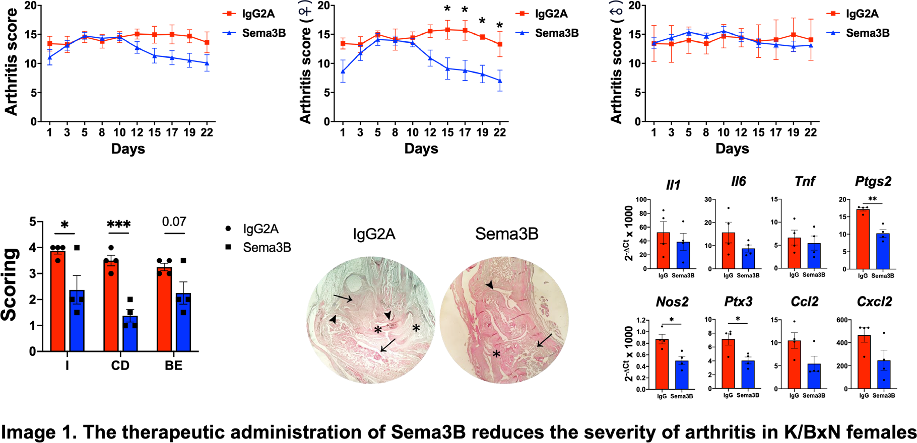
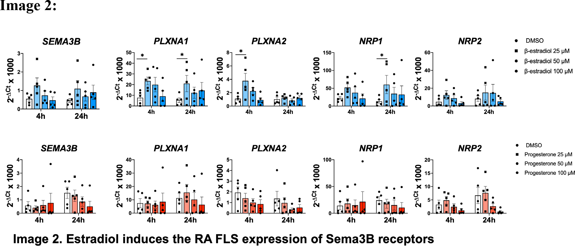
Methods: Recombinant mouse Sema3B or its respective IgG isotype control (both 25 µg/mouse) were administrated in 4 weeks-old K/BxN arthritic mice. Clinical progression and histological analyses were performed. Fibroblast-like synoviocytes (FLS) from patients with RA (all females) were stimulated with increasing concentrations of estradiol and progesterone for 4 and 24h. The mRNA expression in mouse joints and in RA FLS was determined RT-qPCR.
Results: Administration of exogenous Sema3B reduced the clinical severity of arthritis, but only in the female group. Histological analyses of the joints of this group showed a significant reduction in synovial inflammation, cartilage damage, and bone erosion. This effect was associated with a diminished expression of inflammatory mediators, mainly Ptgs2 , Nos2 and Ptx3 . Finally, we found that, in RA FLS, estrogen, but not progesterone, induced the mRNA expression of the Sema3B receptors PlexinA1, PlexinA2 and NRP1.
Conclusions: Therapeutic administration of Sema3B shows a sex-dependent protective effect in a spontaneous model of arthritis. This effect is likely mediated by estrogen-induced activation of Sema3B signaling in rheumatoid arthritis fibroblast-like synoviocytes (RA FLS). These findings suggest that restoring physiological levels of Sema3B may represent a potential therapeutic approach for the treatment of rheumatoid arthritis in women.
REFERENCES: NIL.
Acknowledgments: NIL.
Disclosure of Interests: None declared.