fetching data ... 
Background: Abatacept, an efficacious treatment for rheumatoid arthritis (RA), inhibits the interaction of CD28 on T cells with CD86/80 on antigen-presenting cells. T cells help B cells to produce ACPAs considered pathogenic for RA.
Objectives: The aim of our study was to study the in vivo effect of abatacept on T cell subtypes in RA.
Methods: Twenty-five adult patients with RA (females, 20) not responding to conventional synthetic DMARDs received abatacept and were included in the study. Disease activity as measured by SDAI and DAS28(CRP) was recorded at baseline and at 6-months post treatment. Responders were considered patients who achieved SDAI remission/low disease activity, whereas non-responders patients with SDAI moderate/high disease activity. Serum and peripheral blood samples were collected at baseline and at week 24 post-abatacept treatment. ACPAs were tested using commercial ELISA. Flow cytometry analyses were performed using a FACS Calibur cytometer and the following T cell subtypes were measured at baseline and at 6 months post-treatment: CD4 + T cells, CD8 + T cells, memory/effector CD4 + Tcells(CD45RO + ), naive CD4 + Tcells(CD45RA + ), T peripheral helper cells (PD-1 hi Tph: CD3 + CD4 + PD-1 hi CXCR5 - ), T follicular helper cells (Tfh total: CD3 + CD4 + CXCR5 + CD45RA), ICOS + Tfh, and PD-1 + Tfh. Results are presented as the mean ± SD. Statistical analysis was performed using the 2-tailed t-test and Pearson correlation.
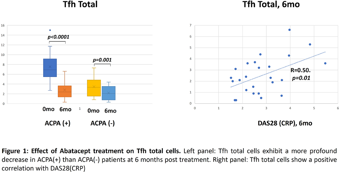
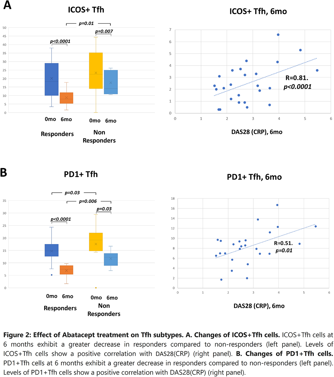
Results: At baseline, ACPA positivity was detected in 52% of patients. Response to abatacept was found in 84.6% ACPA(+) patients and in 58.3% of ACPA(-) patients (p=0.0034). At baseline, PD-1 hi Tph cell and Tfh total cell percentages were higher in ACPA(+) compared to ACPA(-) patients (5.16±2.60 vs 2.80±1.87, p=0.016, and 7.54±3.35 vs 3.45±1.97, p=0.001, respectively) (Figure 1, left panel). Percentages of ICOS + Tfh cells and PD-1+Tfh cells of Tfh cells were lower in responders compared to non-responders (20.1±11.09 vs 26.69±11.05, p=0.014, and 14.73±4.53 vs 20.1±4.89, p=0.030, respectively) (Figure 2A and B left panels). At 6-months, Tfh total cells exhibited a greater decrease in ACPA(+) compared to ACPA(-) patients, as well as ICOS + Tfh cells (47.6±20.98 vs 36.54±27.03, p=0.002) (Figure 1 left panel). Total Tfh cells, ICOS + Tfh cells and PD-1 + Tfh cells exhibited a greater decrease in responders compared to non-responders (Figure 2A and B left panels). DAS28(CRP) score was positively correlated with Tfh total, ICOS + Tfh, and PD-1 + Tfh cells (Figure 1 right panel, Figure 2A and B right panels)
Conclusions: Abatacept was more effective and reduced more deeply Tfh total cells and ICOS+Tfh cells in ACPA(+) than ACPA(-) patients. DAS28(CRP) score was positively correlated with Tfh total cells and Tfh subtypes.
REFERENCES: NIL.
Acknowledgments: NIL.
Disclosure of Interests: Christos Liaskos This study was financed by a BMS grant (IM101-617), Athanasios Mavropoulos This study was financed by a BMS grant (IM101-617), Christina Katsiari This study was financed by a BMS grant (IM101-617), Ioannis Alexiou This study was financed by a BMS grant (IM101-617), Dimitrios Bogdanos This study was financed by a BMS grant (IM101-617), Lazaros Sakkas This study was financed by a BMS grant (IM101-617).