fetching data ... 
Background: CD4 cytotoxic T lymphocytes (CTLs) constitute a heterogeneous T cell population involved in diverse immune responses, ranging from healthy aging to pathological conditions such as autoimmunity and chronic infection [1]. Cytotoxic T cells have traditionally been defined by the expression of effector molecules including granzyme A (GZMA), granzyme B (GZMB), granzyme K (GZMK), perforin (PRF1), and granulysin (GNLY). However, emerging evidence in CD8 T cells indicates that GZMK marks a distinct T cell subset that differs from classical GZMB-expressing cytotoxic T cells and is instead associated with cytokine production and complement activation [2]. In contrast, the functional significance of GZMK-expressing CD4 T cells remain poorly understood. Comprehensive immunophenotyping of CD4 GZMK+ and GZMB+ T cells in rheumatoid arthritis (RA), a prototypical T cell–mediated autoimmune disease, may therefore reveal novel CD4 T cell differentiation states and mechanisms contributing to autoimmune pathology.
Objectives: To identify immunophenotypic features of GZMK+ and GZMB+ CD4 T cells in RA using a multimodal single-cell approach.
Methods: GZMK+ and GZMB+ T cells were identified from an in-house single-cell RNA sequencing (scRNA-seq) dataset of circulating T cells of RA patients (n=3) and healthy donors (HD, n=2) [3] and subjected to downstream analyses. An independent validation cohort was subsequently recruited (RA n=15, HD n=16), in which flow cytometry was performed to assess the expression of phenotypic markers identified in the scRNA-seq data.
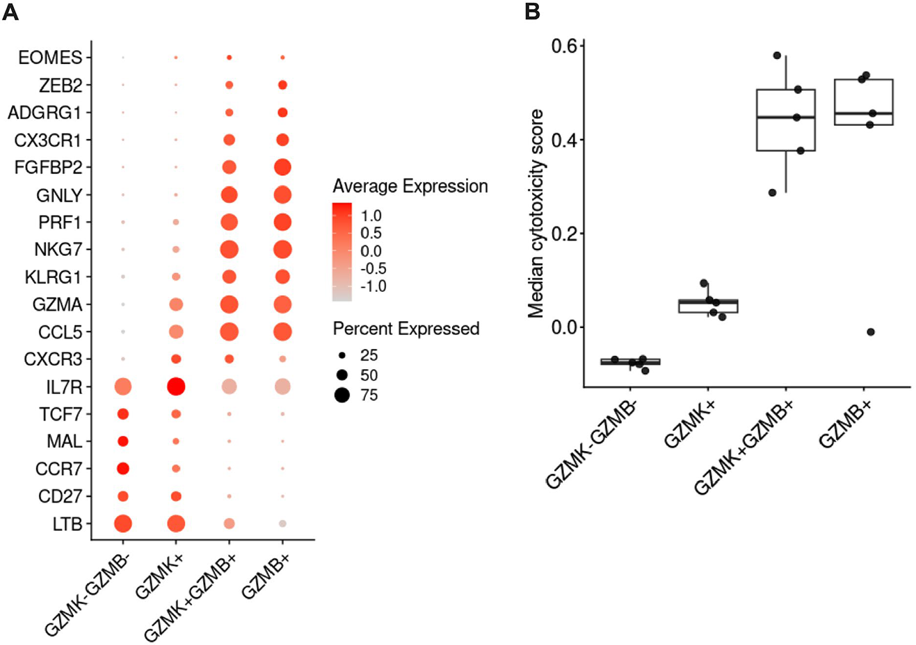
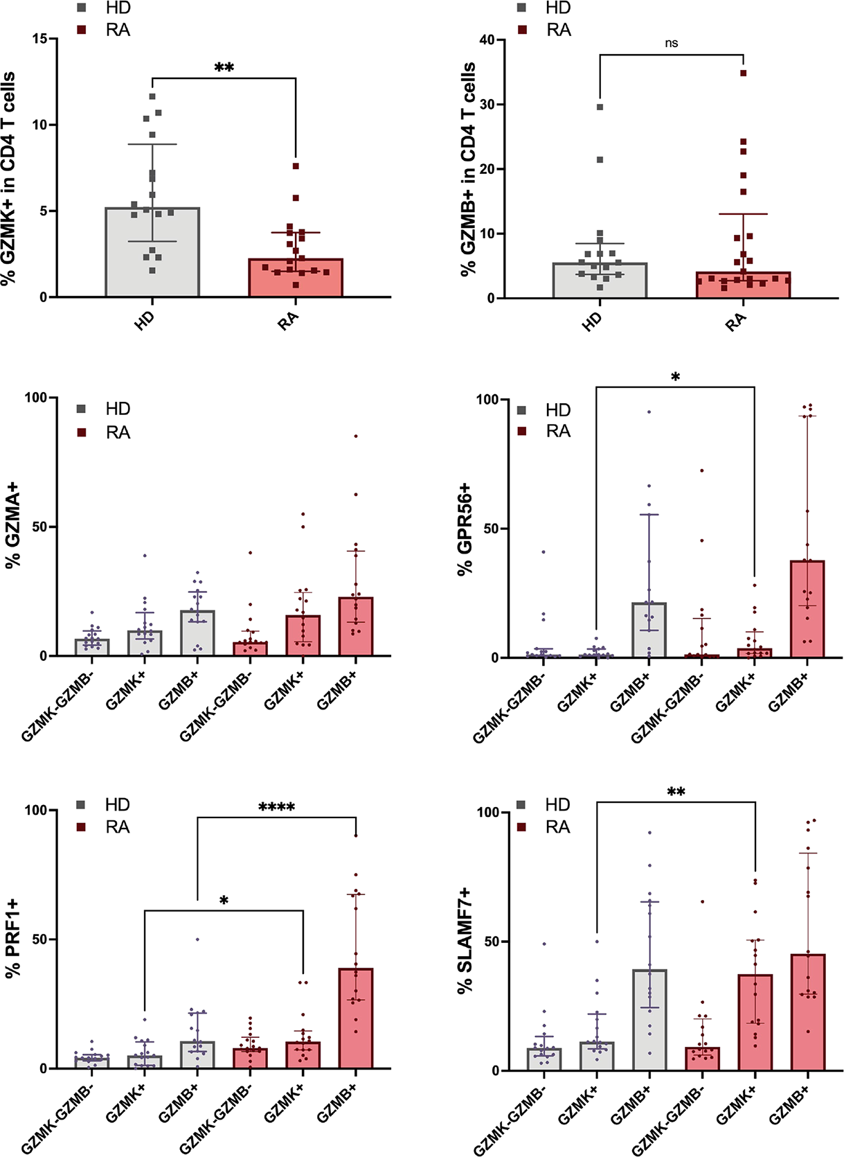
Results: Among all CD4 T cells, 1,707 cells (7.3%) expressed GZMK and 1,509 cells (6.4%) expressed GZMB. Double-positive GZMK+GZMB+ cells were rare (0.5%), whereas the majority were double-negative (GZMK-GZMB-, 85.8%). Differential gene expression analysis showed that GZMK+ CD4 T cells expressed markers associated with lower differentiation states, including CD27, CCR7, and TCF7, while also expressing senescence-associated and cytotoxic markers such as NKG7, KLRG1, GZMA, and PRF1. These cytotoxic markers are characteristic of CD4 CTLs and found enriched in GZMB+ and GZMK+GZMB+ populations. No single marker uniquely defined the GZMK+ CD4 T-cell population. Although IL7R and CXCR3 showed highest expression in GZMK+ cells, both markers were also shared with other CD4 T cell subsets (figure 1A). Cytotoxicity module scores were lowest in GZMK-GZMB- CD4 T cells, intermediate in GZMK+ CD4 T cells, and high in both GZMK+GZMB+ and GZMB+ CD4 T cells (figure 1B). This pattern indicates progressive cytotoxic programming across these populations, with GZMB expression most strongly associated with a cytotoxic transcriptional profile. T cell receptor (TCR) clonality analysis revealed marked clonal expansion within GZMB+ CD4 T cells, which accounted for 53% of all clonally expanded CD4 T cells. Substantial clonal overlap was observed between GZMK+ and GZMB+ populations, with 43.9% of GZMK+ cells and 19.1% of GZMB+ cells sharing TCRs across these subsets, consistent with a shared developmental relationship. Together, these data support GZMK+ CD4 T cells as a distinct differentiation state with an intermediate cytotoxic profile and the capacity for further differentiation into GZMB+ CD4 CTLs. Flow cytometry validation confirmed a gradual increase in cytotoxic marker expression (GZMA, PRF1, GPR56) across GZMK-GZMB-, GZMK+, and GZMB+ CD4 T-cell populations (figure 2). RA patients exhibited similar frequencies of circulating GZMB+ CD4 T cells compared to HD (median 4.2% vs. 5.5%, respectively), but significantly reduced frequencies of GZMK+ CD4 T cells (median 2.3% vs. 5.2%). Notably, GZMK+ CD4 T cells from RA patients expressed higher levels of cytotoxic markers, including GPR56, PRF1 and SLAMF7, than their healthy counterparts. In contrast, the GZMB+ subset was largely comparable between RA and HD, with PRF1 being the only cytotoxic marker upregulated in RA GZMB+ CD4 T cells.
Conclusions: Our data identified GZMK+ CD4 T cells as a distinct differentiation state characterized by partial cytotoxic programming and developmental plasticity toward GZMB+ CD4 CTLs. In RA, this population is reduced in frequency but displays enhanced cytotoxic marker expression, consistent with accelerated cytotoxic maturation of CD4 T cells in autoimmune disease. Altered differentiation dynamics of CD4 CTLs may therefore contribute to RA pathogenesis.
A) Differentially expressed genes in each granzyme subset as indicated. The size of the dots are the percentage of cells expressing a gene, the color reflects the scaled average expression for each gene. B) Median cytotoxicity module score of each granzyme subset. Each point represents one sample, boxplot show median (center line), interquartile range (box) and whiskers extending to 1.5x the interqurartile range.
Flow cytometry measurements of marker expression on CD4 T cells in the validation cohort (RA n=15, HD n=16). The height of the bars indicate the median, whiskers indicate interquartile range.
REFERENCES: [1] Cenerenti M, Saillard M, Romero P, Jandus C. The Era of Cytotoxic CD4 T Cells. Frontiers in Immunology [Internet]. 2022 [cited 2023 Aug 24];13. Available from:
[2] Jonsson AH, Zhang F, Dunlap G, Gomez-Rivas E, Watts GFM, Faust HJ, et al. Granzyme K+ CD8 T cells form a core population in inflamed human tissue. Science Translational Medicine. 2022 June 15;14(649):eabo0686.
[3] Beck F, Nguyen P, Hoffmann A, Loyal L, Thiel A, Melzer M, et al. CD4 + CD8α low T Cell Clonal Expansion Dependent on Costimulation in Patients With Rheumatoid Arthritis. Arthritis & Rheumatology. 2024 Dec;76(12):1719–29.
Acknowledgments: NIL.
Disclosure of Interests: None declared.