fetching data ... 
Background: Immune checkpoint inhibitors have transformed cancer treatment but frequently induce immune-related adverse events. Inflammatory arthritis is among the most common rheumatic toxicities, affecting approximately 5 to 7 percent of treated patients and often resembling rheumatoid arthritis or psoriatic arthritis. Symptoms include persistent joint pain, swelling, and stiffness, frequently requiring long-term immunosuppressive therapy. Unlike classical rheumatoid arthritis, checkpoint inhibitor-induced arthritis often develops in patients without autoantibodies, suggesting distinct immune mechanisms. While synovial inflammation in classical inflammatory arthritis has been well characterized, the immune cell populations driving checkpoint inhibitor-induced disease remain insufficiently defined, particularly across synovial fluid and peripheral blood.
Objectives: To characterize immune cell composition, transcriptional states, and clonal architecture in synovial fluid and peripheral blood from patients with checkpoint inhibitor-induced inflammatory arthritis, and to identify disease-relevant immune clusters for subsequent comparison with classical rheumatoid arthritis and psoriatic arthritis.
Methods: Single-cell RNA sequencing with paired T cell and B cell receptor profiling was performed on CD45 positive cells from synovial fluid mononuclear cells and peripheral blood mononuclear cells obtained from 8 samples collected from 5 patients with checkpoint inhibitor-induced rheumatoid arthritis at arthritis onset. Patients were treated for melanoma, renal, breast, or colorectal cancer. Four patients developed a rheumatoid arthritis-like phenotype and one developed psoriatic-like arthritis. The cohort included 4 ACPA-negative and 1 ACPA-positive individuals. Unsupervised clustering and cell type annotation were conducted using Seurat with reference-based validation approaches. Clonal expansion and tissue sharing of T and B cells between blood and synovial fluid were assessed. Gene regulatory network inference, RNA velocity, cytokine pathways patterns and cell-cell communication analyses are currently under analysis. Comparative analyses with in-house single-cell datasets from rheumatoid arthritis and psoriatic arthritis are ongoing. Key cellular states identified by single-cell RNA sequencing will be validated using multiparameter flow cytometry.
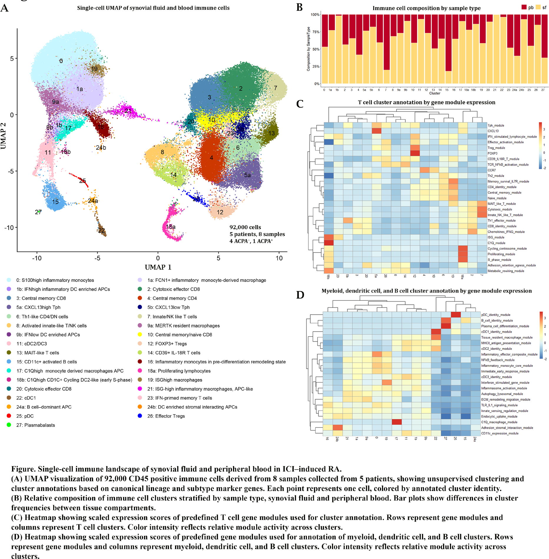
Results: Analysis of approximately 92,000 immune cells identified 33 stable clusters representing two major immune compartments, visualized by unsupervised clustering and annotation of synovial fluid and peripheral blood immune cells (Figure 1). The first compartment comprised myeloid, dendritic cell, and B cell clusters, while the second consisted of multiple transcriptionally distinct T cell clusters. Annotation based on gene module expression defined activated and cytotoxic T cell states, as well as heterogeneous myeloid and antigen-presenting cell populations. Within the myeloid compartment, transcriptional structure revealed two dominant differentiation trajectories emerging from S100high inflammatory monocytes. One trajectory followed a macrophage maturation route, progressing through inflammatory monocyte-derived macrophages into C1Qhigh and highly activated inflammatory macrophage states. A second trajectory showed an interferon-skewed antigen-presenting route, transitioning into dendritic cell-enriched populations, including cDC2 and DC3-like clusters, with evidence of proliferative expansion rather than terminal macrophage differentiation. We identified two subsets of highly cytotoxic effector CD8 + T cells with distinct transcriptional profiles and tissue distributions, one of which was predominantly enriched in synovial fluid and characterized by TIGIT expression Two subsets of peripheral helper T cells (Tph), CXCL13high and CXCL13low, were predominantly derived from synovial fluid, with minor representation in peripheral blood. CXCL13high Tph cells showed prominent representation in the single ACPA-positive patient, consistent with the previous identification of Tph cells in seropositive rheumatoid arthritis. Interestingly, a large B cell cluster predominantly derived from peripheral blood was also primely driven by this single ACPA-positive patient. In general, several clusters were highly abundant in synovial fluid compared with peripheral blood and displayed transcriptomic signatures suggestive of disease relevance. These differentiation patterns will be further investigated using RNA velocity and trajectory analyses and validated by flow cytometry.
Conclusions: Checkpoint inhibitor-induced inflammatory arthritis displays a distinct immune architecture characterized by synovial enrichment of specific immune clusters and divergent myeloid differentiation programs. Integrated single-cell profiling with planned flow cytometric validation will help understand the immune mechanisms driving arthritis following ICI-therapy and inform improved treatment approaches for this emerging group of arthritis.
REFERENCES: NIL.
Acknowledgments: NIL.
Disclosure of Interests: None declared.