fetching data ... 
Background: Sarcoidosis is a heterogenous granulomatous disease, in which Interferon γ (IFNγ) and Tumour Necrosis Factor α (TNFα) are key immunological mediators. The disease is associated with increased risk of malignancy. Anti-cytokine autoantibodies (aabs) have emerged as important modulators of immune responses in autoimmune diseases, yet their presence and relevance in sarcoidosis remain unexplored.
Objectives: This study investigated IFN aabs in sarcoidosis and their potential association with disease heterogeneity and malignancy risk.
Methods: Serum samples from 167 Icelandic patients with biopsy-proven sarcoidosis (collected 2009–2013) were analysed for aabs against type I–III IFNs, including assessment of neutralising capacity, using a magnetic multiplex particle-based assay and Reporter HEK-Blue cells, respectively. Clinical data, including malignancy diagnoses, were retrieved from medical records. Autoantibody profiles were compared with those of systemic lupus erythematosus (SLE) patients and healthy controls.
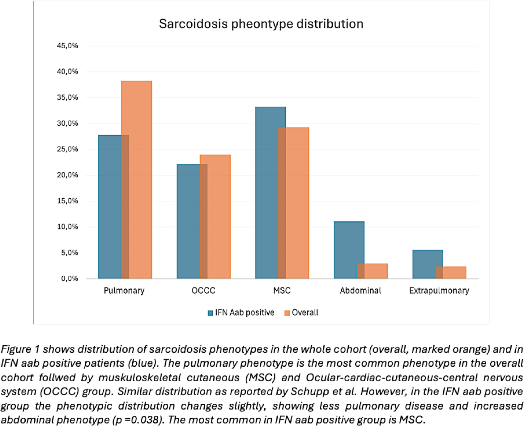
Results: Eighteen sarcoidosis patients (10.8%) tested positive for IFN Aabs. (Table 1) Two patients had neutralising aabs. IFN aab positivity was associated with a fourfold increased risk of malignancy (OR 3.99, 95% CI 1.40–11.36; p=0.009). Smoking was not a significant predictor of malignancy after sarcoidosis diagnosis (OR 0.977, 95% CI 0.43-2.18; p=0.954). Compared with SLE and healthy controls, sarcoidosis patients exhibited increased aab responses against type I and II IFNs, with a predominance of the IgG isotype. Clinically, IFN aab-positive patients showed a shift from typical pulmonary involvement toward increased abdominal involvement (p=0.038). (Table 1, Figure 1)
Conclusions: IFN autoantibodies are present in a subset of sarcoidosis patients and are associated with increased risk of malignancy. IFN aab positive patients exhibit clinical features with a shift towards increased abdominal involvement. These findings suggest a potential role for IFN aabs in sarcoidosis immunopathogenesis and highlight the need for further prospective studies.
Overview sarcoidosis patients, overall, positive and negative IFN autoantibodies (aabs), phenotypes and symptoms
| IFN aab positive
| IFN aab negative (89.2%, n=149) | Total (n=167) | Test (p-value) | |
|---|---|---|---|---|
| Sex (female ) | 13 (72.2%) | 70 (47.0%) | 83 (49.7%) | 0.043 |
| Age mean (years ) | 45.939 (12.934) | 44.624 (12.216) | 44.766 (12.262) | 0.669 |
| History of smoking | 14 (77.8%) | 77 (51.7%) | 91 (54.5%) | 0.036 |
| Malignancy | 8 (44.4%) | 25 (16.8%) | 33 (19.7%) | 0.009 |
| Phenotype | ||||
| Pulmonary | 5 (27.8%) | 59 (41.3%) | 64 (39.8%) | 0.271 |
| OCCC | 36 (25.2%) | 4 (22.2%) | 40 (24.8%) | 0.785 |
| MSC | 6 (33.3%) | 43 (30.1%) | 49 (30.4%) | 0.777 |
| Abdominal | 2 (11.1%) | 3 (2.1%) | 5 (3.1%) | 0.038 |
| Extrapulmonary | 1 (5.6%) | 3 (2.1%) | 4 (2.5%) | 0.374 |
| Respiratory symptoms | ||||
| Dyspnea | 6 (33.3%) | 56 (39.7%) | 62 (39.0%) | 0.601 |
| Mucous | 2 (11.1%) | 22 (15.6%) | 24 (15.1%) | 0.616 |
| Pleuritic chest pain | 5 (27.8%) | 31 (22.0%) | 36 (22.6%) | 0.580 |
| Cough | 4 (22.2%) | 56 (39.4%) | 60 (37.5%) | 0.155 |
| Treatment | ||||
| No treatment | 4 (22.2%) | 35 (23.8%) | 39 (23.6%) | 0.881 |
| Treatment | 14 (77.8%) | 97 (66.0%) | 111 (67.3%) | 0.314 |
| Non-responders | 0 (0.0%) | 7 (4.8%) | 7 (4.2%) | 0.344 |
| No information treatment | 0 (0.0%) | 17 (11.4%) | 17 (10.2%) | |
Table 1 shows overview of the sarcoidosis patients; those positive and negative for IFN aabs as well as the whole cohort. Patients were categorized into one of five described by Schupp et al. OCCC: Ocular-cardiac-cutaneous-central nervous system phenotype. MSC: muskuloskeletal-cutaneous phenotype. The patients were additionally categorized according to the treatment they received, as an indicator of disease severity, burden and impact of life. Treatment: corticosteroids and/or disease modifying agents. Resistant: those who needed treatment with TNF-α inhibitors. Statistically significant difference was seen in ratio of female patients in the IFN aab positive group as well as history of smoking, malignancy and abdominal phenotype.
Distribution of sarcoidosis phenotypes in the whole cohort and IFN autoantibody (aab) positive patients
REFERENCES: NIL.
Acknowledgments: NIL.
Disclosure of Interests: None declared.