fetching data ... 
Background: Among the various chemokines implicated in lupus nephritis (LN) pathogenesis, interferon-γ-inducible protein 10 (IP-10) is a major mediator that recruits immune cells and induces inflammation. Secreted IP-10 promotes the migration of CD8 + T cells and NK cells to the inflammatory site, inducing an inflammatory response in the kidneys and causing tissue damage. T stem-like cells have been shown to differentiate into terminally exhausted T-cells in chronic infection models when IP-10 levels increase or persist during chronic inflammation. However, it remains unclear whether CD8 + T-cells differentiate into the exhausted lineage and if IP-10 influences T-cell exhaustion in LN.
Objectives: This study aims to determine the role of IP-10 in the reduction of T stem-like cells and accumulation of terminally exhausted T-cells in LN.
Methods: Proteome profiler screening was performed on plasma samples obtained from patients with SLE (n=6), LN (n=6), and healthy controls (n=6). In MRL/lpr mice, serum was acquired bi-weekly from weeks 10 to 24, and the levels of serum anti-dsDNA, cytokines (BAFF, IL-16, CD30, Leptin, TIM-3, and VEGF), and chemokines (IP-10, MCP-1, MIG, and I-TAC) were measured using ELISA and Luminex assays. To evaluate CD8 + T cell differentiation in tissues, the spleen and kidney tissues was obtained and analyzed by flow cytometry at 10, 14, 18, and 24 weeks of age.
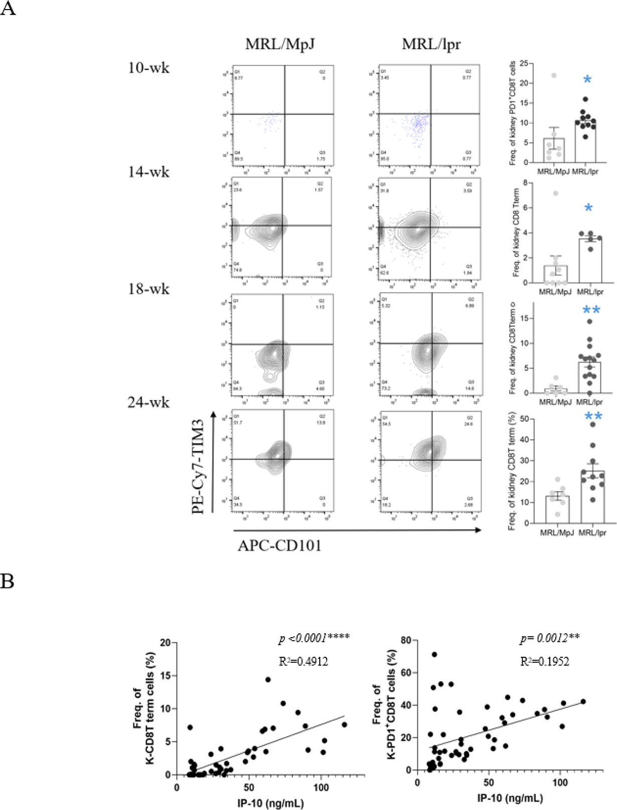
Results: Among the 105 circulating factors analyzed, IP-10 demonstrated the highest level of elevation in patients with SLE and LN compared to healthy controls. Correlation analysis of these factors revealed that in patients with LN, IP-10 was strongly associated with other immunoregulatory factors, such as MCP-1 and TIM-3. In the MRL/lpr mouse model, serum IP-10 levels increased with age and correlated with BAFF and anti-dsDNA levels. Additionally, flow cytometry analysis of spleen from MRL/lpr mice revealed that T stem-like cells (TCF1 + CX3CR1 + CD8 + ) within the tissue decreased and differentiated into an exhausted state, leading to the accumulation of terminally exhausted T-cells (TCF1 - CX3CR1 - CD8 + ) correlating with serum IP-10 levels. In the kidneys of MRL/lpr mice, terminally exhausted T-cells and IP-10 levels increased with age, showing a significant association over time (Figure 1).
Conclusions: The accumulation of terminally exhausted T-cells in the kidney was associated with the level of serum IP-10 in lupus prone mice, suggesting an interaction between IP-10 and the cells within the LN.
Accumulation of terminally exhausted T cells in the kidney (A) and their correlation with serum IP-10 levels (B) in MRL/lpr LN mice. ****p < 0.0001, ***p < 0.001, **p < 0.01, *p < 0.05 vs. control (Mann-Whitney U test).
REFERENCES: [1] Anders HJ, Saxena R, Zhao MH, Parodis I, Salmon JE, Mohan C. Lupus nephritis. Nat Rev Dis Primers. 2020;6(1):7.
[2] Puapatanakul P, Chansritrakul S, Susantitaphong P, Ueaphongsukkit T, Eiam-Ong S, Praditpornsilpa K, et al. Interferon-Inducible Protein 10 and Disease Activity in Systemic Lupus Erythematosus and Lupus Nephritis: A Systematic Review and Meta-Analysis. Int J Mol Sci. 2019;20(19).
[3] Ozga AJ, Chow MT, Lopes ME, Servis RL, Di Pilato M, Dehio P, et al. CXCL10 chemokine regulates heterogeneity of the CD8(+) T cell response and viral set point during chronic infection. Immunity. 2022;55(1):82–97 e8.
[4] Zhang Q, Shan Y, Shen L, Ni Q, Wang D, Wen X, et al. Renal remodeling by CXCL10-CXCR3 axis-recruited mesenchymal stem cells and subsequent IL4I1 secretion in lupus nephritis. Signal Transduct Target Ther. 2024;9(1):325.
[5] Wu C, Jiang S, Chen Z, Li T, Gu X, Dai M, et al. Single-cell transcriptomics reveal potent extrafollicular B cell response linked with granzyme K(+) CD8 + T-cell activation in lupus kidney. Ann Rheum Dis. 2024.
Acknowledgments: NIL.
Disclosure of Interests: None declared.