fetching data ... 
Background: Systemic lupus erythematosus (SLE) is preceded by a prolonged preclinical phase characterized by progressive immune dysregulation. Although T and NK cells dysregulation persists in established SLE, their roles in the earliest stages of disease pathogenesis remain poorly elucidated.
Objectives: This study aimed to comprehensively investigate the phenotypic and functional alterations in T and NK cell populations throughout SLE development.
Methods: Peripheral blood samples were collected from three cohorts, namely healthy controls (HC, n = 4), individuals with preclinical SLE (pre-SLE, n = 4), and patients with newly diagnosed SLE (n = 5). Peripheral blood mononuclear cells (PBMCs) were isolated via density gradient centrifugation, followed by single-cell RNA sequencing (scRNA-seq) to delineate the transcriptional landscapes of T and NK cells at single-cell resolution, and single-cell T cell receptor sequencing (scTCR-seq) to investigate the clonal repertoire.
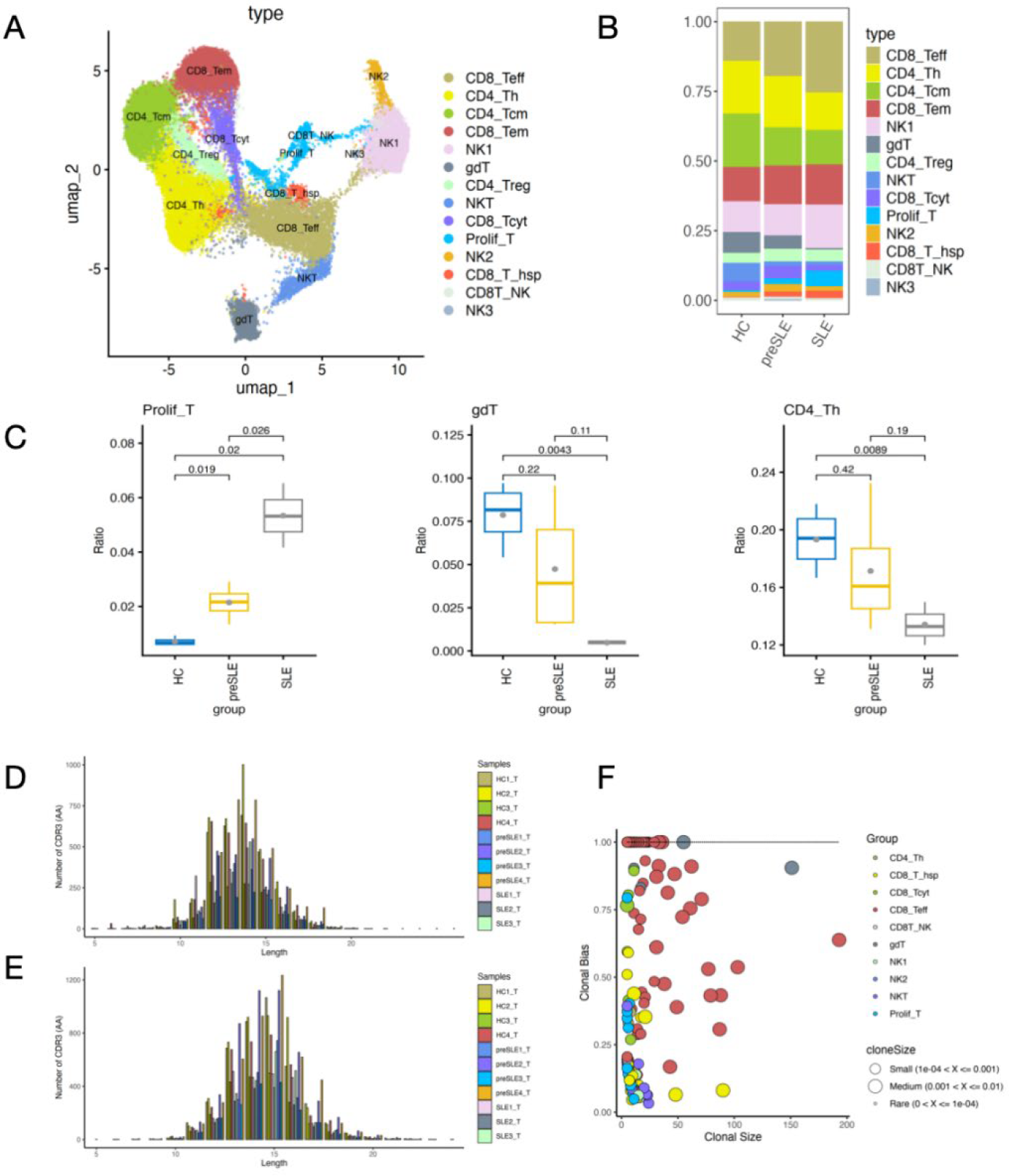
Results: A total of over 100,000 cells were captured for profiling, with 11 T-cell subpopulations and 3 NKT cell subset identified. The frequency of proliferating T cells (prolif_T) showed a significant stepwise increased from HC to pre-SLE to SLE ( P < 0.05 for all pairwise comparisons). Conversely, γδT (gdT) and CD4 + T helper (CD4_Th) cells proportions decreased in SLE, with significant differences only between the SLE and the HC group ( P = 0.043 and 0.0089, respectively). TCRα/β chains lengths were concentrated within 10-20 bp; CD8 + effector T(CD8_Teff) and gdT cells exhibited marked clonal expansion bias, and the proportion of unique T cell clones gradually decline with SLE progression.
Conclusions: These findings provide novel insights into the dynamic immunological alterations of T and NK cells during SLE development, shedding light on potential early biomarkers and pathogenic mechanisms of SLE.
Dynamic shifts in T and NK cell populations and clonality during SLE progression. (A) UMAP visualization of T and NK cell subpopulations. (B) The relative proportions of T cell (n = 11) and NK cell (n = 3) subpopulations across HC (n = 4), pre-SLE (n = 4), and SLE (n = 5). (C) Ratios of Prolif_T, gdT, and CD4_Th were compared across the three groups. Length distributions of TCRα (D) and TCRβ (E) chains. (F) Clonal bias of T/NK cell subsets.
REFERENCES: NIL.
Acknowledgments: NIL.
Disclosure of Interests: None declared.