fetching data ... 
Background: Systemic Lupus Erythematosus (SLE) is a chronic, multisystem autoimmune disease that predominantly affects women of reproductive age and remains a major cause of mortality in this group. It is characterized by the production of diverse autoantibodies and systemic immune dysregulation. Emerging evidence highlights significant metabolic reprogramming in immune cells as a key feature of SLE pathogenesis. Recent studies have demonstrated that nuclear receptor corepressors NCoR1 and SMRT regulate immune cell metabolism, particularly in dendritic cells, and influence downstream T-cell responses. Their altered expression correlates with inflammatory profiles in autoimmune diseases like RA. Understanding these metabolic alterations may offer valuable insights into SLE prognosis and guide the development of personalised therapeutic approaches. Based on these concepts, we examined the expression of NCoR1 and SMRT in different immune cells, along with glycolytic activity in T cells, in SLE, RA and healthy controls.
Objectives: This study aimed to quantify NCoR1 and SMRT protein levels in immune cells from SLE, RA, and healthy controls and to evaluate glycolytic activity in T cell subsets to explore their potential role in SLE pathogenesis.
Methods: This was a cross-sectional, observational, single-centre study involving 80 SLE patients meeting the 2019 ACR/EULAR classification criteria along with 40 RA patients (ACR/EULAR, 2010) and 50 age-matched healthy controls. Clinical data, physical examination findings, and routine laboratory investigations were recorded. SLE disease activity was assessed using the SLEDAI-2K score. 25-colour FACS using Cytek Aurora (spectral flowcytometry) was defined for profiling all the immune cells, including DCs (CD141 & CD1c), macrophages, monocytes, different subtypes of T-cells (CD3, CD4, CD8, memory CD4 & CD8, effector Th1, Th2, Th17, and TFH cells). NCoR1 and SMRT protein levels were quantified in immune cells via flow cytometry, and their ratio was calculated and analysed in relation to disease activity and treatment regimens in SLE patients. Glycolytic activity in T cell subsets was assessed using the SCENITH protocol. Descriptive statistics were reported as means and standard deviations. NCoR1 and SMRT expression data were visualized using the ggplot2 package in R. Correlations with SLEDAI scores were evaluated using Pearson’s correlation via the ggpubr package. Between-group differences were analyzed using unpaired Student’s t-tests, with p-values ≤ 0.05 considered statistically significant.
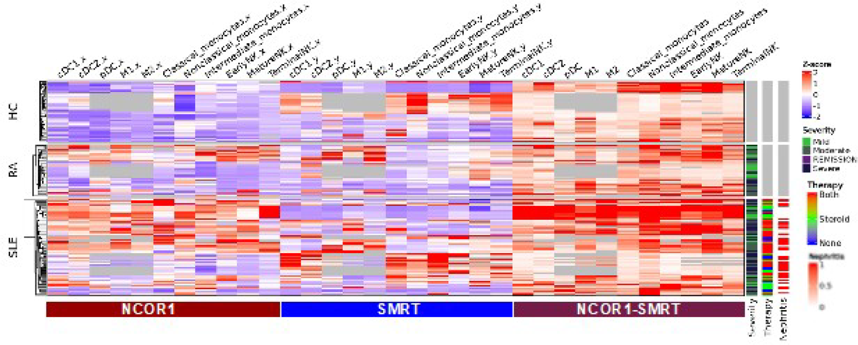
Results: SLE patients (mean age 27.1 ± 8.7) were younger than RA (39.4 ± 8.9) and healthy controls (32.4 ± 8.6). Majority of the patients in the current cohort had mucocutaneous (87.5%), renal (62.5%), and joint involvement (60%), respectively, with a mean baseline SLEDAI of 10.5. Most patients (45%) were on both steroids and immunosuppressants, 35% on steroids alone, and 20% received only hydroxychloroquine monotherapy at the time of enrolment. Immune profiling revealed significantly increased NCoR1 expression in dendritic cells, macrophages, and NK cells in both RA and SLE compared to HC. SMRT expression was elevated in cDC2, non-classical/intermediate monocytes (SLE), and pDC, M2 macrophages (RA), but downregulated in monocytes and cDC1 (RA). SLE patients exhibited a significantly higher NCoR1/SMRT ratio in cDC1, pDC, M1/M2 macrophages, non-classical monocytes, and NK cells. Various T cell subsets like Tfh, Th1, Th17, activated CD4 + , CD4 + TEM and activated CD8 + T cells showed a marked increase in the NCoR1/SMRT ratio in SLE patients (p < 0.0001). However, there was a negative correlation with SLEDAI and absence of impact of treatment on NCoR/SMRT ratio. Notably, SLE showed significantly elevated glycolytic flux in T cell subsets except CD4 + central memory, Tfh, or Treg cells. Glycolysis was particularly elevated in activated CD4 + T cells in moderate to severe SLE patients as compared to mild cases.
Conclusions: This study is the first of its kind to profile NCoR1 and SMRT protein expression across a broad range of immune cell subsets in SLE patients. The altered NCoR1/SMRT ratio observed in key innate and adaptive cell populations, particularly in activated T cells, underscores a potential immunoregulatory axis relevant to the pathogenesis of SLE. These findings provide novel perspectives on transcriptional and metabolic disruptions in lupus, which could lead to future treatments targeting immune cell metabolism and nuclear receptor signalling pathways.
Heat map showing expressions of NCoR1, SMRT and NCoR1/SMRT ratio across innate immune cells among the study population
REFERENCES: NIL.
Acknowledgments: NIL.
Disclosure of Interests: None declared.