fetching data ... 
Background: Systemic lupus erythematosus (SLE) is characterized by profound T cell dysregulation, particularly involving aberrant CD4 + T cell activation and differentiation. Immune checkpoint receptors have emerged as key regulators of T cell fate decisions beyond classical activation or exhaustion paradigms. T cell immunoglobulin and mucin domain–containing protein 3 (TIM-3) is implicated in immune regulation across autoimmune and inflammatory conditions; however, its role in shaping CD4 + T cell functional states during early SLE remains poorly understood.
Objectives: To define TIM-3–associated phenotypic and functional heterogeneity of CD4 + T cell subsets in early SLE and to explore their immunological relevance.
Methods: Peripheral blood mononuclear cells from patients with early-stage SLE and healthy controls were analyzed using multiparameter flow cytometry. CD4 + T cells, including regulatory T cells (CD3 + CD4 + FoxP3 + ), were stratified by TIM-3 expression and differentiation status using CD45RA and CCR7. Expression of functional and regulatory markers, including granzyme B, CD39, HLA-DR, CD226, CD62L, and vanin-2, was assessed. Associations between TIM-3–defined subsets and clinical parameters were evaluated.
Results: TIM-3 expression delineated distinct CD4 + T cell states in early SLE. TIM-3 + regulatory T cells exhibited a redistribution toward resting phenotypes with a relative reduction in activated regulatory subsets, suggesting altered regulatory dynamics. Among conventional CD4 + T cells, TIM-3 expression was preferentially associated with terminally differentiated effector memory populations and was accompanied by increased expression of activation and migratory markers. Notably, both TIM-3 + regulatory and effector CD4 + T cells co-expressed granzyme B and CD39, indicating a hybrid cytotoxic–regulatory functional profile. In early SLE, TIM-3 expression was aberrantly expanded within naïve and central memory CD4 + T cell compartments, reflecting disturbed T cell differentiation trajectories.
Conclusions: TIM-3 marks a distinct functional state of CD4 + T cells in early SLE characterized by combined cytotoxic and immunoregulatory features. These findings highlight TIM-3 as a key immune checkpoint associated with altered CD4 + T cell fate and immune imbalance in autoimmune disease.
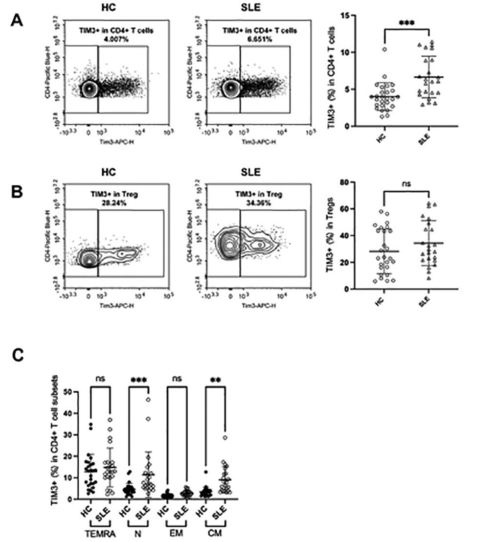
Comparison of expression of TIM3 in CD4 T cell and Treg subsets between healthy individuals and SLE patients. Peripheral blood mononuclear cells (PBMCs) were extracted from healthy individuals and SLE patients.A&B. Representative scatter plots of cells analyzed by flow cytometry. Comparison of the percentages of TIM3+ in CD4+ T cells(in A) and Tregs(in B) between HCs and SLE patients. C. Comparison of the percentages of TIM3+ in TEMRA,N,EM,CM in CD4+ T cell subsets between HC individuals and SLE patients, using ANOVA test for comparisons among multiple groups. Each symbol represents the data of a person. Data are presented as mean with SD. Ns not significance,** P < 0.01,*** P < 0.001.
REFERENCES: NIL.
Acknowledgments: NIL.
Disclosure of Interests: None declared.