fetching data ... 
Background: Vascular Adhesion Protein-1 (VAP-1) is an endothelial adhesion molecule with enzymatic activity that mediates leukocyte trafficking and vascular inflammation [1]. Its pathophysiological role in systemic vasculitides, particularly Giant Cell Arteritis (GCA) [2] remains poorly defined. This study investigated VAP-1 expression in patients with GCA and analyzed the inflammation-dependent regulation of endothelial VAP-1, including the modulatory effects of glucocorticoids.
Objectives: This research aimed to determine the clinical relevance of Vascular Adhesion Protein-1 in Giant Cell Arteritis. The study investigated the induction of this adhesion molecule under inflammatory conditions and assessed the capacity of glococorticoid to suppress its expression in both macrovascular and microvascular endothelial models.
Methods: Patients with GCA and age- and sex-matched control subjects were recruited for a cross-sectional study. VAP-1 mRNA expression was determined from peripheral blood samples using quantitative real-time polymerase chain reaction (qPCR). Human aortic endothelial cells (HAoEC) and human umbilical vein endothelial cells (HUVEC) were cultured and stimulated with pro-inflammatory mediators, including tumor necrosis factor (TNF), interferon-gamma (IFN-ϒ), and lipopolysaccharide, at concentrations of 10 and 100 ng/ml at time points of 12 and 24 hours. VAP-1 expression was quantified using flow cytometry with a standardized gating strategy. The modulating effect of prednisolone on cytokine-induced VAP-1 expression was statistically evaluated in analog stimulation assays.
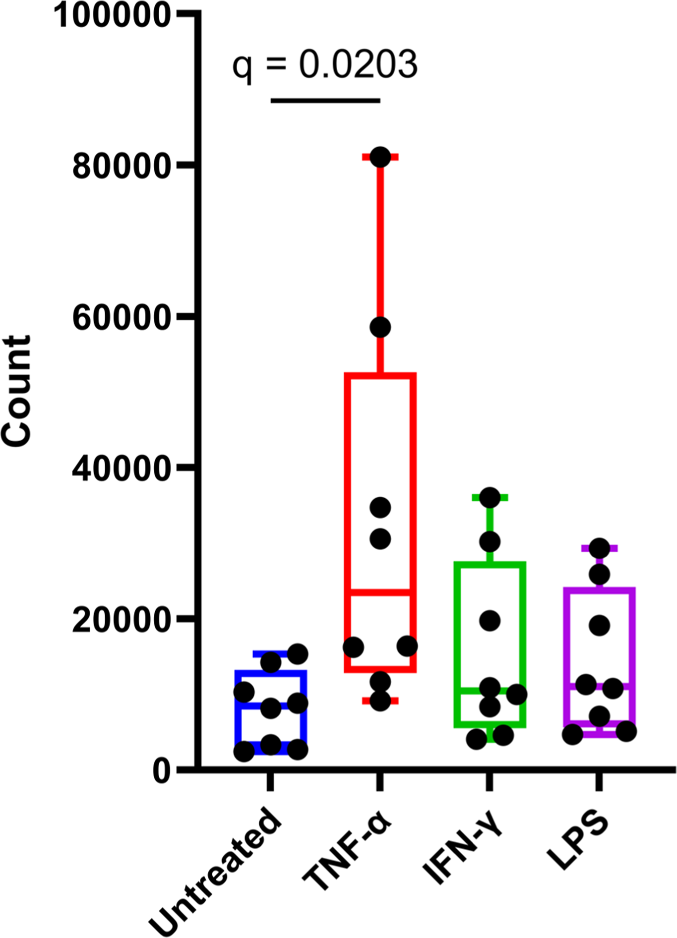
Results: Analysis of patient samples showed a significant upregulation of VAP-1 mRNA in patients with GCA compared to controls, with a mean fold change of 33.8 ± 10.1 versus 1.00 ± 0.08 (p = 0.048). Stimulation of endothelial cells with pro-inflammatory mediators led to a marked induction of VAP-1 expression. TNF showed the strongest effect, at a concentration of 100 ng/ml, it resulted in a proportion of approximately 80 to 90 percent VAP-1 positive cells (p ≤ 0.02, Figure 1). IFN-γ and lipopolysaccharide also induced significant upregulation of VAP-1 expression, though to a lesser extent than TNF. Concentration-dependent effects were observed for all stimuli. Conversely, treatment with prednisolone led to a clear attenuation of cytokine-induced VAP-1 expression. Prednisolone significantly reduced VAP-1 levels in cells stimulated by TNF, IFN-γ, and lipopolysaccharide, with consistently negative effect estimates across all conditions. A comparison between aortic and umbilical vein endothelial cells revealed no significant differences in VAP-1 expression under basal or stimulated conditions.
Conclusions: This study demonstrates for the first time that VAP-1 is significantly upregulated in patients with GCA. Furthermore, endothelial VAP-1 expression is strongly induced by pro-inflammatory stimuli and effectively suppressed by glucocorticoid therapy. The results support a functional role for VAP-1 in the vascular inflammatory response and highlight its potential value as a biomarker and therapeutic target in vasculitides such as Giant Cell Arteritis.
This figure displays the frequency of cells positive for Vascular Adhesion Protein-1 across different inflammatory states. Compared to the control, stimulation with tumor necrosis factor-alpha (q ≤ 0.02), interferon-gamma, and lipopolysaccharide resulted in a significant elevation of protein expression.
REFERENCES: [1] Petzinna SM, Küppers J, Schemmer B, Kernder AL, Bauer CJ, Baerlecken NT, et al. Advanced imaging of relapse in giant cell arteritis: The role of vascular adhesion protein-1 and [68Ga]Ga-DOTA-Siglec-9 positron emission tomography-computed tomography. J Intern Med. 2025 June 26;298(2):138–42.
[2] Petzinna SM, Küppers J, Schemmer B, Kernder AL, Bauer CJ, von der Emde L, et al. Case report: Detecting giant cell arteritis in [68Ga]Ga-DOTA-Siglec-9-PET/CT. Front Immunol. 2024;15:1501790.
Acknowledgments: NIL.
Disclosure of Interests: None declared.