fetching data ... 
Background: Gout is a systemic metabolic disease characterized by chronic urate burden and frequent renal involvement. Gouty nephropathy and urate-associated metabolic chronic kidney disease (CKD) often develop subclinically and therefore remain under-recognized in routine practice. Conventional renal ultrasound is operator-dependent and insufficiently standardized for detecting early metabolic and crystal-associated renal changes. Bubnov’s patented ultrasound methodology (UA 66903) [1] integrates structural, vascular, and biomechanical renal features to identify metabolic and urate-related nephropathy; however, its complexity limits reproducibility across operators. Artificial intelligence (AI) may support standardization while preserving expert-driven interpretation.
Objectives: To evaluate the feasibility of AI-assisted renal ultrasound analysis for detecting gouty nephropathy according to the integrated ultrasound framework, with human expert control over anatomical sign validation and final diagnosis.
Methods: We analyzed 50 renal ultrasound images acquired according to the patented ultrasound framework [1], which defines diagnostic sonographic features of gouty nephropathy using integrated B-mode, intrarenal Doppler, and shear-wave elastography (SWE) criteria [2]. Renal ultrasound examinations were performed in patients with hyperuricemia and gout. Image evaluation assessed predefined anatomical signs, including cortical echogenicity and microgranularity, corticomedullary differentiation, renal contour, subcapsular hypoechoic striae, parenchymal microcalcifications, intrarenal resistive index (RI), and SWE stiffness distribution.
An AI-based extension was applied to quantify image features, highlight candidate pathological signs, and estimate pattern probabilities. Each detected sign and the overall classification were verified or corrected by an expert operator. Differentiation from other metabolic CKD patterns (diabetic, hypertensive, and obesity-related) was performed using combined structural, vascular, and SWE criteria.
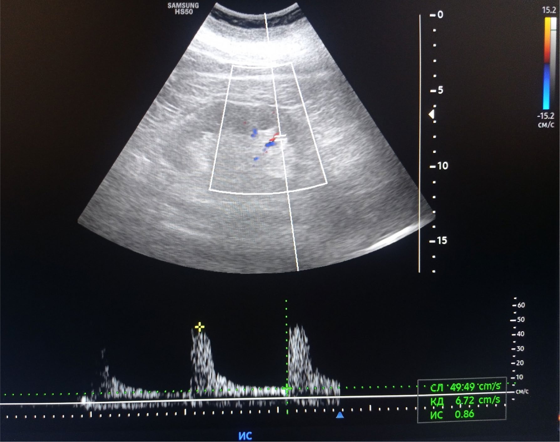
Results: AI reliably identified and quantified key ultrasound features associated with urate-related renal injury, including increased cortical echogenicity with microgranularity, impaired corticomedullary differentiation, elevated RI, and heterogeneous SWE stiffness. Human–AI synthesis improved consistency of sign recognition and supported classification of gouty nephropathy as a distinct metabolic CKD pattern (figure 1). Expert oversight remained essential for accurate anatomical interpretation and final diagnosis, particularly in patients with mixed metabolic phenotypes [3].
Conclusions: AI-assisted ultrasound analysis, applied within the ultrasound framework for urate-associated nephropathy, enhances standardization and reproducibility of renal ultrasound assessment in gout. This human-controlled AI approach facilitates early detection and differentiation of gouty nephropathy within the spectrum of metabolic CKD, supporting precision management of patients with hyperuricemia and gout.
AI-assisted ultrasound assessment of gouty nephropathy.
Renal B-mode ultrasound demonstrates increased cortical echogenicity with heterogeneous microgranular texture, impaired corticomedullary differentiation, irregular renal contour, subcapsular hypoechoic striae, and focal parenchymal microcalcifications. Color and spectral Doppler evaluation reveals elevated intrarenal resistive index (RI), indicating combined metabolic and vascular involvement. Artificial intelligence (AI)–based image analysis supports detection and quantification of pathological features, including echogenicity distribution, textural heterogeneity, and vascular indices. Final interpretation is performed under expert supervision, integrating anatomical validation and shear-wave elastography (SWE)–derived stiffness patterns to distinguish urate-associated metabolic nephropathy from other causes of chronic kidney disease.
REFERENCES: [1] Bubnov RV. Method for diagnosis of gouty nephropathy. Patent UA66903U, Ukraine; 25 Jan 2012. Available from:
[2] Bubnov R. Shear wave elastography effective in ultrasound diagnosis of chronic kidney disease in patients with hyperuricaemia and gout. Nephrol Dial Transplant. 2022; 37(Supplement_3):gfac096.004..
[3] Bubnov R. Ultrasound imaging patterns of kidney disease in patients with metabolic syndrome. Nephrol Dial Transplant, 2023; 38(Supplement_1):gfad 063c_4869.
Acknowledgments: NIL.
Disclosure of Interests: None declared.