fetching data ... 
Background: MDA5 + dermatomyositis (MDA5 + DM) frequently complicates interstitial lung disease (ILD), especially rapidly progressive ILD (RP-ILD), which worsens prognosis. Traditional assessments cannot directly reflect fibroblast activation status, a key driver of pulmonary fibrosis. 68Ga-FAPI-04 PET/CT targets fibroblast activation protein (FAP), but its value in MDA5 + DM remains understudied.
Objectives: Investigate the capacity of this technology in reflecting pathological changes and analyze its quantitative results’ correlation with clinical characteristics such as lung function to explore the potential application value of this technique in predicting the progression of lung lesions in patients with MDA5 + DM.
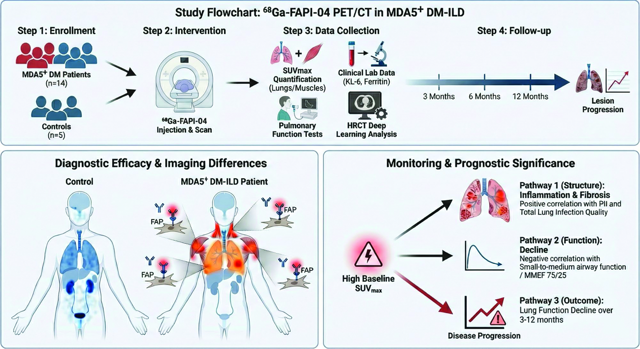
Methods: Fourteen MDA5 + DM patients and 5 controls were enrolled. 68Ga-FAPI-04 PET/CT was performed to obtain SUVmax of lungs, muscles, and other organs. Clinical data, pulmonary function tests, and deep learning-quantified HRCT indices (e.g., PII) were collected. Follow-ups (3/6/12 months) for pulmonary function and HRCT were conducted. Correlations between SUVmax, clinical features, and follow-up outcomes were analyzed (figure 1).
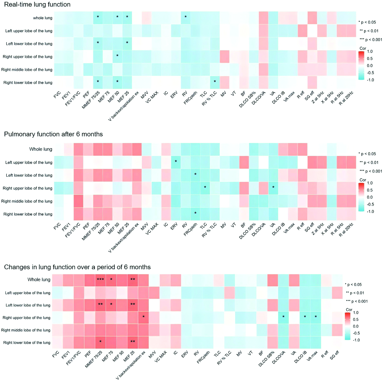
Results: Whole-lung SUVmax was significantly higher in MDA5 + DM patients (3.32 vs 0.67, p<0.0001), with left lower lobe showing the most prominent difference (p=0.0003). SUVmax strongly correlated with HRCT inflammation/fibrosis indices (e.g., left lower lobe vs PII at 3 months: r=0.9863, p<0.0001) and small-to-medium airway function (left upper lobe vs MMEF 75/25: r=-0.8168, p=0.0133). Baseline SUVmax predicted 3–12-month lesion progression and lung function changes, with deceased patients having higher baseline whole-lung SUVmax (6.21±0.85 vs 3.24±1.72, p=0.008) (figure 2).
Conclusions: 68Ga-FAPI-04 PET/CT sensitively reflects fibroblast activation, assesses early pulmonary impairments, and predicts long-term progression in MDA5 + DM, providing a valuable tool for clinical monitoring and intervention.
Study design, diagnostic imaging patterns, and clinical utility of 68Ga-FAPI-04 PET/CT in MDA5 + DM-ILD. The top panel outlines the study workflow. The bottom left panel demonstrates diagnostic efficacy. The bottom right panel highlights monitoring value. MDA5 + DM, anti-Melanoma Differentiation-Associated gene 5 positive dermatomyositis; ILD, Interstitial Lung Disease; SUVmax, Maximum Standardized Uptake Value; HRCT, High-Resolution Computed Tomography; FAP, Fibroblast Activation Protein; PII, Pulmonary Inflammation Index; MMEF, Maximal Mid-expiratory Flow.
These heatmaps illustrate the correlations between 68Ga-FAPI-04 PET/CT-derived SUVmax (across whole lung, left upper/lower lobes, right upper/middle/lower lobes) and pulmonary function indices, including real-time function, function at 6 months, and percentage changes in function over 6 months. The color scale represents Pearson correlation coefficients (Cor), with red indicating positive correlations and blue indicating negative correlations. Statistical significance is denoted as * p < 0.05, ** p < 0.01, *** p < 0.001.
REFERENCES: NIL.
Acknowledgments: NIL.
Disclosure of Interests: None declared.