fetching data ... 
Background: Tendon stiffness reflects the mechanical integrity of tendon tissue and is fundamental for effective functional performance, distributing mechanical load, and protection against injury. Age-related changes in collagen structure, hormonal influences, body composition, and mechanical loading from physical activity have all been linked to alterations in tendon mechanical behaviour; however, the existing evidence remains inconsistent. The associations between tendon stiffness and age, sex, body mass index (BMI), and physical activity are not yet well understood, and comparative data across upper and lower limb tendons are limited. Shear wave elastography (SWE) is an emerging ultrasound technique that enables quantitative assessment of tendon stiffness and offers an opportunity to determine how these potential confounding factors are associated with tendon stiffness across different tendon sites.
Objectives: To investigate the association between tendon stiffness, measured using shear wave elastography, and potential confounding factors including age, sex, BMI, and physical activity level across the supraspinatus tendon (SST), common extensor tendon (CET), patellar tendon (PT), and Achilles tendon (AT).
Methods: This observational cross-sectional study was conducted at the Leeds Biomedical Research Centre, Chapel Allerton Hospital, Leeds, with data collected from July 2024 to September 2025. Ethical approval was obtained (ref: 23/PR/1001), and written informed consent was secured from all participants. SWE was used to quantify tendon stiffness, expressed as mean shear wave velocity (m/s), with a 15–6 MHz matrix linear transducer. SWE measurements were obtained from mid-tendon regions during a single session. Three SWE readings were recorded per tendon, and mean values were used for analysis. The region of interest (ROI) size was set to 2.5 mm 2 , with measurements obtained in the longitudinal plane using a stand-off gel layer applied to the skin surface. Demographic and clinical variables collected included age (years), sex, BMI (kg/m 2 ), and physical activity level. Physical activity was categorised as high, moderate, or low according to the International Physical Activity Questionnaire (IPAQ). High activity was defined as ≥3 days of vigorous activity or 7 days of combined activity achieving ≥1500–3000 MET-min/week; moderate activity included ≥5 days of combined activity achieving ≥600 MET-min/week or walking for at least 30 minutes per day; low activity included participants not meeting these criteria. Descriptive statistics were used to summarise demographic data. Multiple linear regression models were constructed separately for each tendon to assess independent associations with tendon stiffness. Regression coefficients (β) with corresponding 95% confidence intervals were reported to describe the direction and magnitude of associations. Model fit was assessed using adjusted R 2 values, and statistical significance was set at p < 0.05.
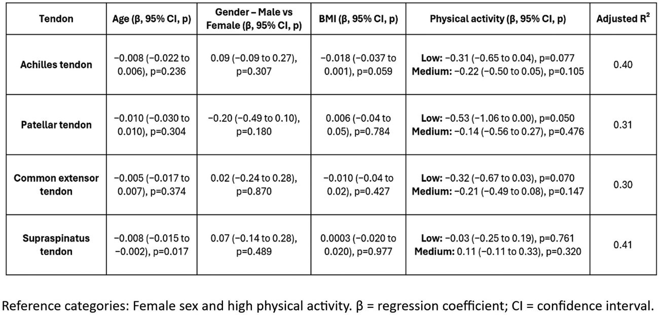
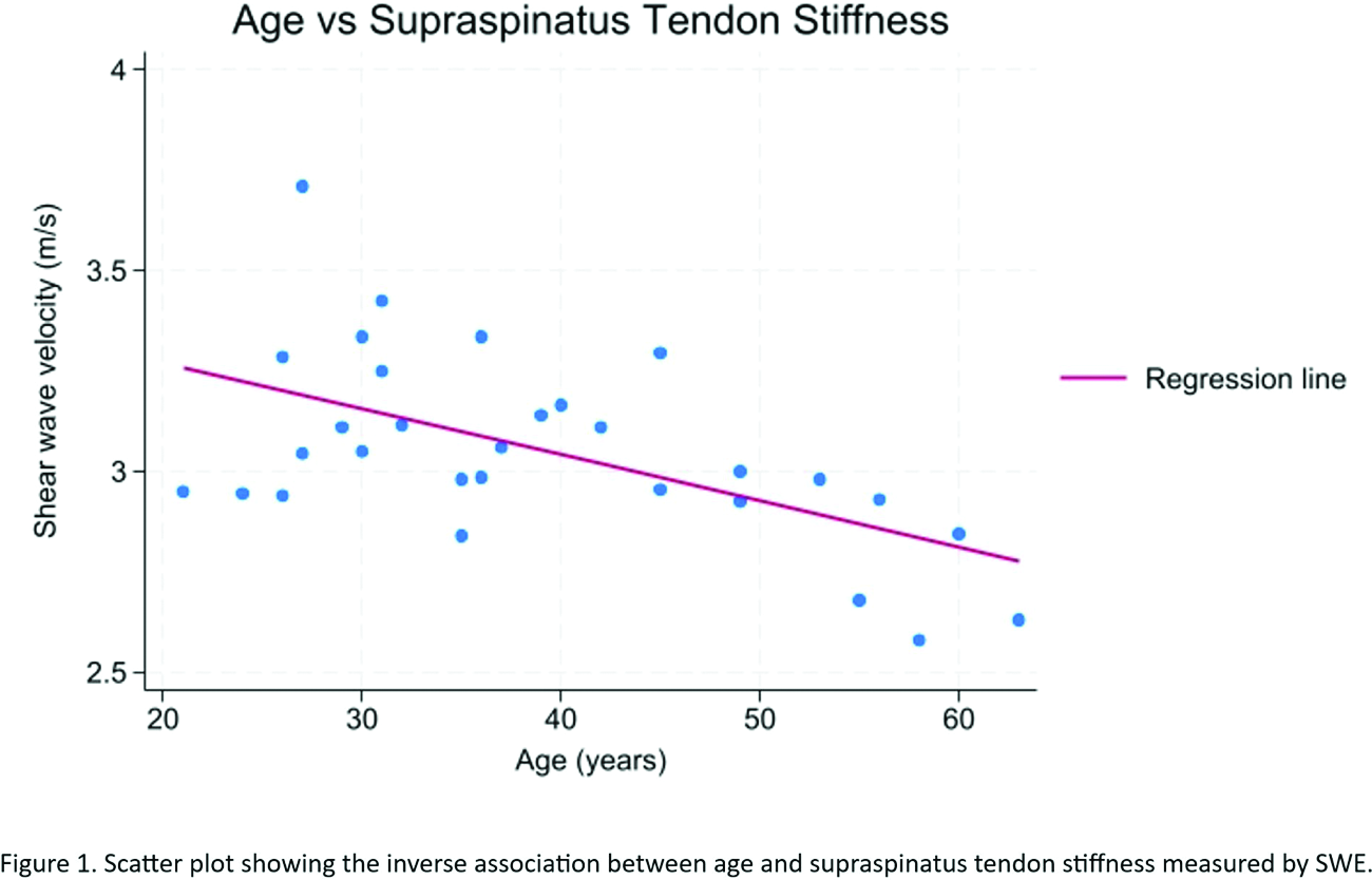
Results: Thirty asymptomatic adult volunteers (12 males and 18 females; mean age ± SD 38.9 ± 11.85; range 21–63 years; mean BMI ± SD 24.02 ± 3.92 kg/m 2 ), participated in the study. Multiple linear regression was used to examine the associations between age, sex, BMI, physical activity level, and tendon stiffness. Overall, model fit was moderate, with adjusted R 2 values ranging from 0.30 to 0.41 across tendons (Table 1). Age was significantly associated with supraspinatus tendon stiffness, with increasing age associated with lower stiffness (β = -0.008 m/s per year; p = 0.017). This finding may reflect age-related tendon degeneration and reduced collagen within the rotator cuff. Although negative associations between age and stiffness were also observed for the common extensor, patellar, and Achilles tendons, these relationships were not statistically significant (β = -0.005 m/s per year, p = 0.374; β = -0.010 m/s per year, p = 0.304; and β = =0.008 m/s per year, p = 0.236, respectively). Males demonstrated slightly higher stiffness values than females; however, these differences were small and not statistically significant across all examined tendons, indicating that sex was not an independent predictor of tendon stiffness. BMI was not significantly associated with tendon stiffness across tendons, although a borderline inverse association was observed for the Achilles tendon, higher BMI associated with lower stiffness (β = -0.018 m/s per kg/m 2 ; p = 0.059). Physical activity level was associated with patellar tendon stiffness, with lower activity levels linked to reduced stiffness compared with high activity (β = -0.53; p = 0.05). No significant associations between activity level and stiffness were observed for the other tendons, although trends towards lower stiffness with reduced activity were noticed.
Conclusions: In this multi-tendon shear wave elastography study, age was negatively associated with tendon stiffness, although this association was statistically significant only for the supraspinatus tendon, which showed a clear decrease in stiffness with increasing age. The absence of significant sex- and BMI-related differences may reflect the relatively small sample size, which limits the power to detect modest differences. Lower physical activity was significantly associated with reduced patellar tendon stiffness. Overall, these findings indicate that age and physical activity are important determinants of tendon stiffness in specific tendons. Further studies in larger and more diverse populations are needed to confirm these findings.
Table 1. Multiple linear regression analysis of age, sex, BMI, and physical activity in relation to tendon stiffness by SWE.
REFERENCES: NIL.
Acknowledgments: NIL.
Disclosure of Interests: None declared.