fetching data ... 
Background: Musculoskeletal ultrasound (US) enables dynamic visualization of muscle architecture and trigger points (TrPs), yet conventional interpretation remains qualitative. The fractal analysis method (UA Patent 63457, 2011) [1,2] quantifies biological texture complexity using fractal dimension (FD) and lacunarity, reflecting tissue micro-organization. Integration with ultrasound-guided dry needling (DN-US) allows objective monitoring of structural normalization in myofascial pain syndromes.
Objectives: To apply AI-assisted 3D fractal analysis to B-mode and shear-wave ultrasound images of the lumbar region (multifidus MF, quadratus lumborum QL, thoracolumbar fascia TLF) and quantify pre-/post-DN structural changes in acute and chronic low-back-pain (LBP) patients.
Methods: Thirty LBP patients (16 female; mean age 42 ± 9 yrs) underwent DN-US therapy following Bubnov’s visualized TrP technique [3-5]. Imaging: 2D and 3D B-mode, SWE (Samsung HS50). ROIs: MF, QL, TLF; hypoechoic TrPs and needle paths identified in multiplanar reconstructions. Processing: AI-guided segmentation (U-Net) with subsequent fractal analysis (box-counting and multifractal spectrum) per Bubnov method; parameters FD and lacunarity Λ were computed for each ROI. Validation: Phantoms and repeated scans ensured inter-rater reliability (ICC > 0.9). FD values were correlated with SWE stiffness and clinical status (acute < 2 weeks; chronic > 2 months).
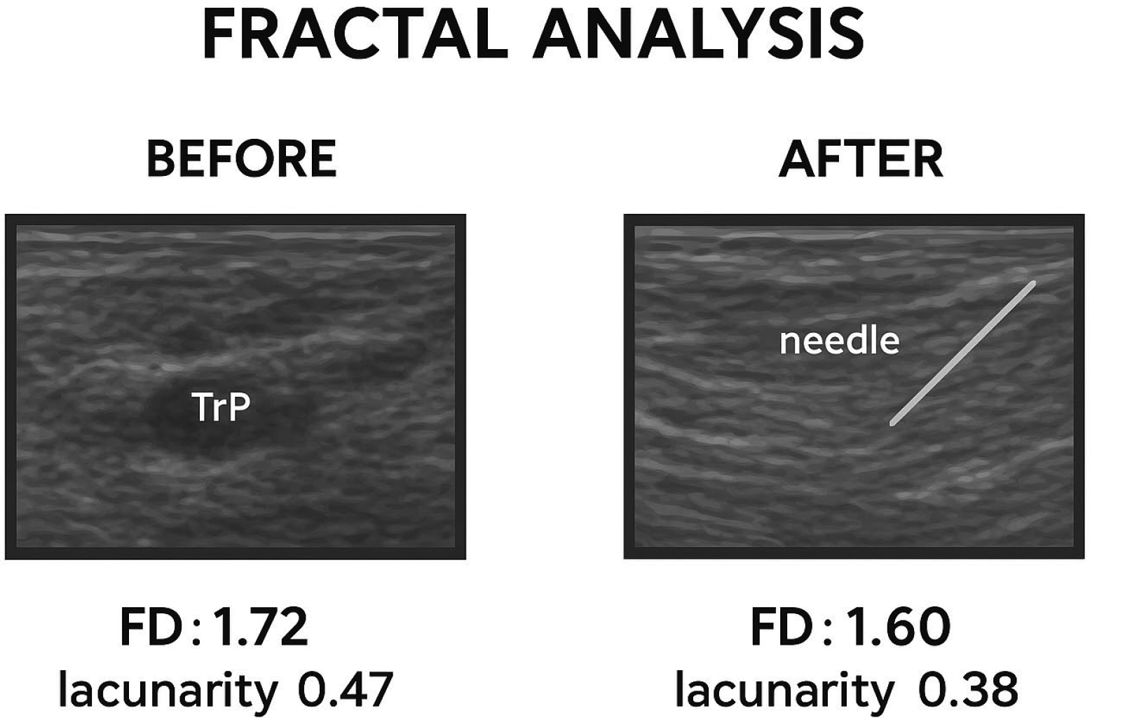
Results: Trigger points (TrPs): Acute LBP: small (2–5 mm) active TrPs with SWE 20–27 kPa; moderate fractal irregularity ( FD 1.52 ± 0.04 , Λ 0.34). Chronic LBP: larger, hypovascular, fibrotic TrPs with SWE 5–8 kPa; pronounced fractal disorganization ( FD 1.72 ± 0.05 , Λ 0.47). Post-DN: FD decreased by 0.06 ± 0.02 and Λ by 0.05, indicating restoration of ordered fascicular alignment. Thoracolumbar fascia (TLF): Chronic cases showed thickened (≥ 5 mm) hyperechoic fascia with high heterogeneity ( FD 1.25–1.30 , Λ 0.55). Acute cases exhibited near-normal linear structure ( FD 1.10–1.15 , Λ 0.32). Quadratus lumborum (QL) and multifidus (MF): Chronic LBP: spastic/atrophic texture, blurred fascicles ( FD 1.70 ± 0.05 ). Acute LBP: hypertonic but organized ( FD 1.55 ± 0.04 ). Post-DN: FD normalized toward 1.50 and fascicular contrast improved on 3D rendering. 3D multiplanar findings: Fractal analysis of 3D volumes confirmed increased surface complexity in chronic fibrosis ( surface FD ≈ 2.35 vs 2.15 normal ) and localized normalization after DN.
Conclusions: AI-assisted ultrasound fractal analysis provides a reproducible, objective biomarker of paraspinal muscle and fascial organization in low back pain. Trigger point activity is characterized by increased fractal complexity, which normalizes following effective ultrasound-guided dry needling. This approach extends prior applications of fractal analysis in muscle spasticity to LBP and offers a quantitative bridge between imaging, biomechanics, and clinical pain assessment, supporting precision diagnostics and personalized interventional strategies.
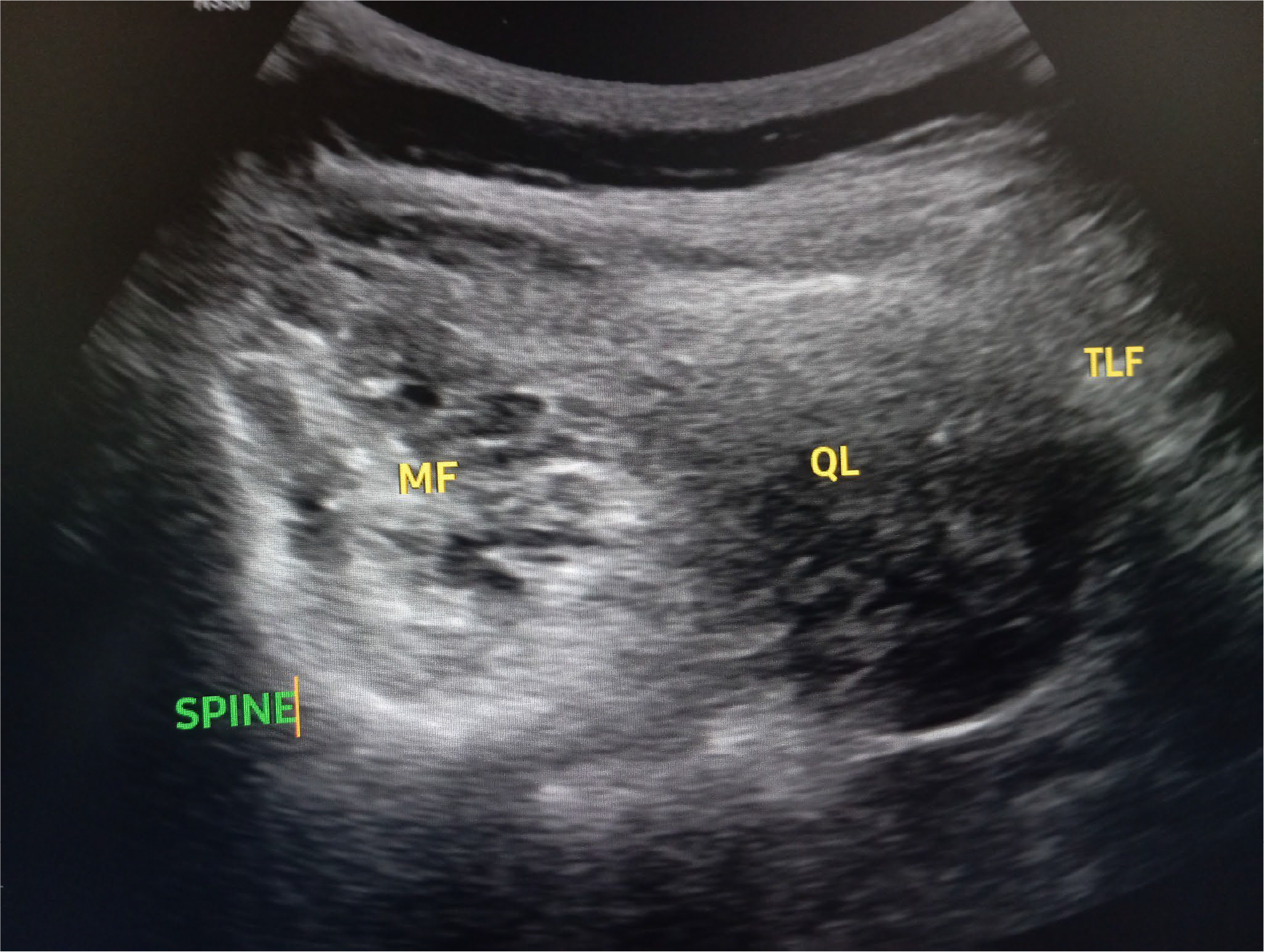
High-quality transverse lumbar ultrasound image of the paraspinal compartment with AI-assisted segmentation of multifidus, quadratus lumborum, and thoracolumbar fascia, illustrating regions of interest for fractal analysis.
AI-based fractal analysis according to Bubnov’s patented method quantitatively distinguishes acute vs chronic LBP by capturing micro-architectural complexity of muscle and fascia.
• Acute pattern: high stiffness but moderate FD → reversible spasticity.
• Chronic pattern: low stiffness, high FD and Λ → fibrotic disorganization.
• Therapeutic response: post-DN reduction in FD (≈ 0.05–0.10) signifies restored hierarchical muscle order.
This integrative 3D-multiplanar fractal-DN-US approach provides an objective biomarker framework for personalized pain diagnostics and monitoring of rehabilitation outcomes.
REFERENCES: [1] Patent No. 790559 u 2011 02941 Ukraine. Method of fractal analysis of medical images/Bubnov R. V., Melnik I. M. – Published 14.03.2011.
[2] Bubnov RV, Melnyk IM. The methods of fractal analysis of diagnostic images. initial clinical experience. Lik Sprava. 2011 Apr-Jun;(3-4):108-13.
[3] Bubnov RV. Evidence-based pain management: is the concept of integrative medicine applicable? EPMA J 2012, 3(1):13.
[4] Bubnov, R.V. FRI0541 Trigger Points Dry Needling Under Ultrasound Guidance for Low Back Pain Therapy. Comparative Study. Ann. Rheum. Dis. 2015, 74, 624.
[5] Bubnov R, Kalika L. THE ROLE OF THORACOLUMBAR FASCIA ULTRASOUND IN LOW BACK PAIN - IMPLICATION FOR GUIDED DRY NEEDLING. Annals of the Rheumatic Diseases 2022;81:1714.
Acknowledgments: NIL.
Disclosure of Interests: None declared.