fetching data ... 
Background: Takayasu arteritis (TAK) is a large-vessel vasculitis primarily affecting the aorta and its branches, usually in young adults. Musculoskeletal involvement in TAK has not been systematically characterized; while spondyloarthritis-like peripheral features have been reported [1], a polymyalgia rheumatica (PMR)-like pattern remains unclear. FDG-PET/CT enables semi-quantitative assessment of subclinical musculoskeletal inflammation and may clarify these patterns.
Objectives: To investigate the prevalence and distribution of musculoskeletal involvement in TAK using PET/CT compared with age- and sex-matched controls, and to explore whether a PMR-like pattern is associated with older age.
Methods: We retrospectively analyzed 95 PET/CT scans of clinically active TAK patients fulfilling ACR-1990 criteria. Musculoskeletal regions (joints, entheses, trochanteric, interspinous ligaments) were assessed semi-quantitatively using SUVmax and SUVmean. Lesion-to-liver ratios >1 defined positivity. Vascular activity was quantified by PETVAS. Controls were 49 individuals with negative work-up for lung masses.
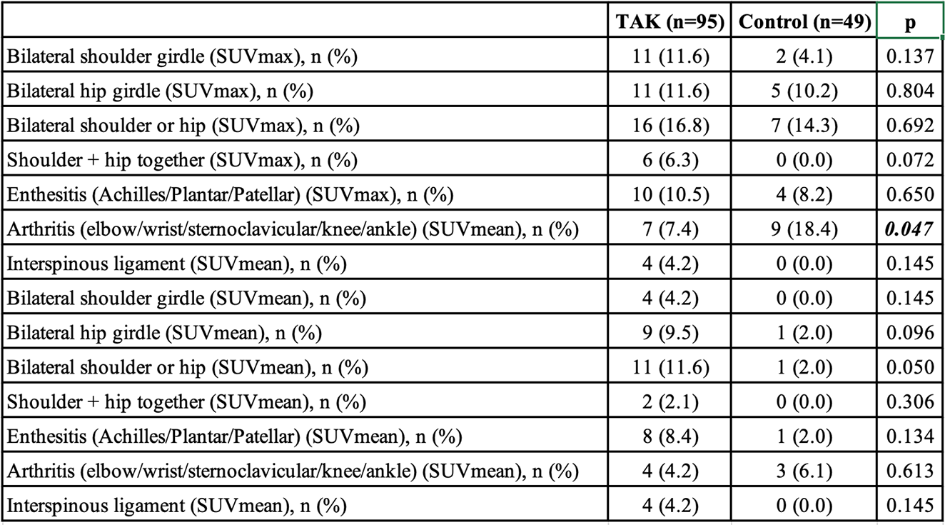
Results: Patients (mean age 38.7±13.8; 81F/14M) had mean PETVAS 3.9±7.2. No clinical arthritis, enthesitis or bursitis was observed at imaging. Vascular uptake was most frequent in the aortic arch (16.8%). Enthesitis rates were comparable between groups (p=0.650). Arthritis was more frequent in controls by SUVmax (p=0.047), whereas SUVmean suggested higher prevalence in TAK. Bilateral shoulder and combined shoulder–hip uptake tended to be higher in TAK, without statistical significance (Table-1). Subclinical arthritis was associated with older age in TAK (62.4±8.9 vs. 47.6±12.9 years, p=0.004). PMR-like shoulder–hip patterns were age-related, while enthesitis tended to occur in younger patients. Figure 1 provides an illustrative example of TAK and PMR overlap.
Conclusions: Subclinical musculoskeletal FDG uptake was generally more frequent in TAK, with PMR-like distributions emerging in older age and enthesitis in younger patients. These findings suggest that, in older individuals, TAK may display GCA/PMR-like musculoskeletal features. Larger cohorts and standardized imaging protocols are warranted.
FDG-PET/CT findings consistent with Takayasu arteritis.
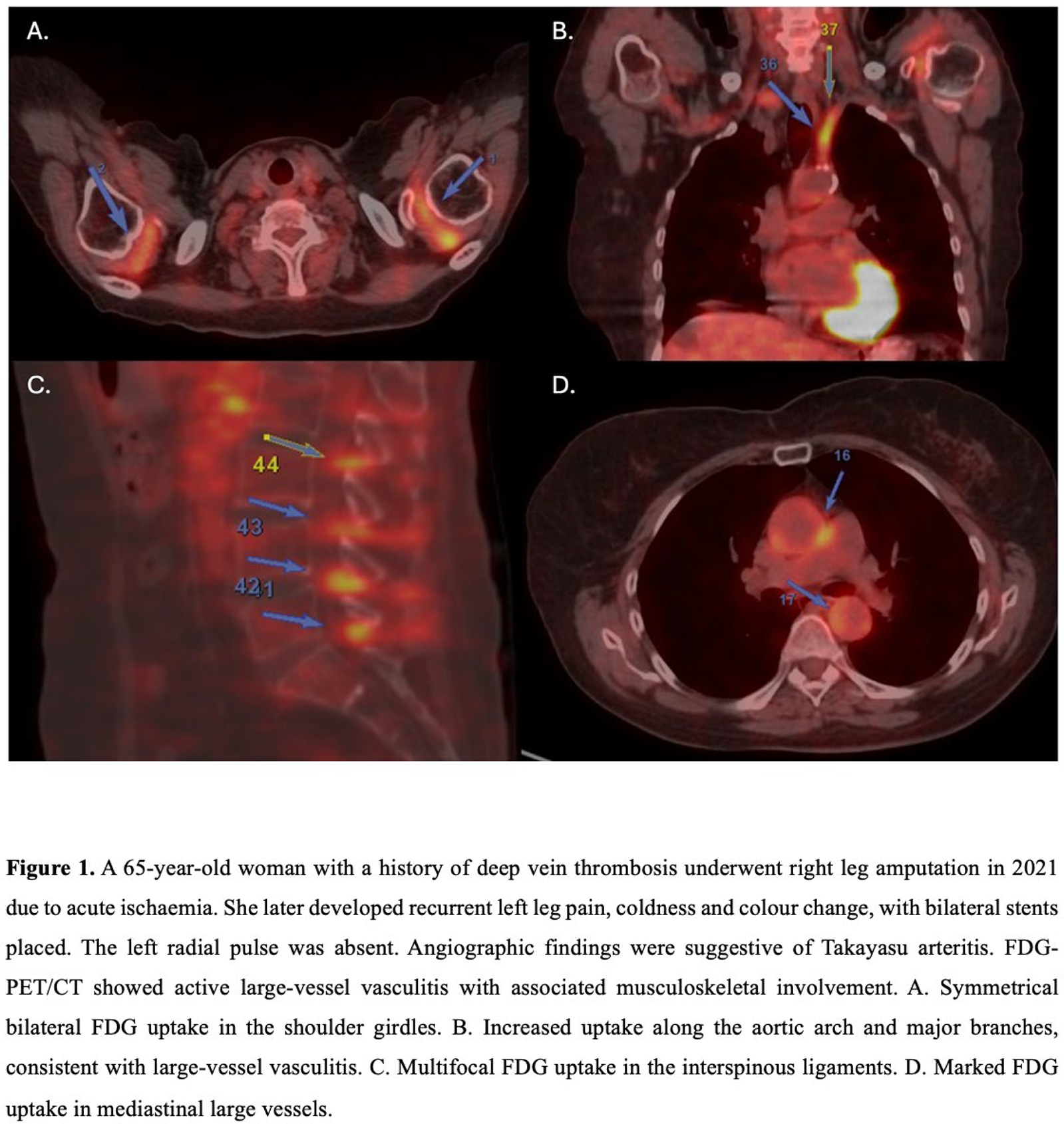
Table 1. Musculoskeletal involvement patterns in Takayasu arteritis compared with controls
REFERENCES: [1] Abacar K, et al. Clin Rheumatol. 2024 May;43(5):1571-1578.
Acknowledgments: NIL.
Disclosure of Interests: None declared.