fetching data ... 
Background: Sjögren’s disease (SjD) is a systemic autoimmune condition affecting exocrine glands, leading to progressive glandular dysfunction and, in some cases, ectopic lymphoid structure (ELS) formation. Salivary gland epithelial cells (SGECs) actively contribute to disease pathogenesis, yet the mechanisms linking epithelial stress to persistent inflammation remain poorly understood. Our studies indicate that SGECs from SjD patients display altered autophagy and metabolic activity, suggesting that epithelial energy homeostasis may be tightly linked to inflammatory chronicity.
Objectives: To define how metabolic pathways are remodeled during salivary gland inflammation and to determine whether epithelial metabolic dysfunction in salivary glands is associated with inflammation severity and epithelial activation in SjD.
Methods: Metabolic remodeling was investigated in vivo using a murine model of virus-induced ELS formation in salivary glands based on intraductal adenoviral cannulation. Transcriptomic profiling of whole glands and isolated SGECs was performed at day 0, day 12 and day 20 post-cannulation, followed by pathway-level analyses. Human minor salivary gland biopsies from SjD patients and sicca controls were analyzed by bulk transcriptomics and histopathology, with stratification by ELS status. Primary human SGECs were studied by mass-spectrometry-based metabolomics, Seahorse assays of glycolysis (ECAR) and oxidative phosphorylation (OCR), and integrated transcriptomic and proteomic profiling. To dissect metabolic alterations, SGECs were treated in vitro with agents affecting inflammatory activation (poly(I:C)), glycolysis (2-DG) and OXPHOS (oligomycin). Single-cell RNA sequencing was used to identify epithelial subtypes with altered metabolic activity.
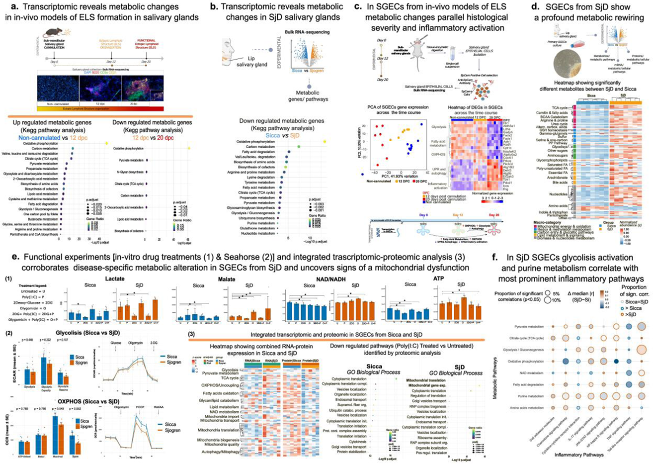
Results: In murine salivary glands, ELS formation was associated with progressive metabolic remodeling. At peak inflammation (day 12), glands exhibited marked downregulation of OXPHOS, accompanied by alterations in pyruvate metabolism and the tricarboxylic acid (TCA) cycle. These changes were consistent with a shift toward alternative energy-producing pathways, including glycolysis (Figure 1a). Concordantly, transcriptomic profiling of human salivary glands identified distinct metabolic signatures separating SjD patients from sicca controls, characterized by impaired OXPHOS and engagement of alternative bioenergetic programs (Figure 1b). These metabolic alterations correlated with histological severity, with ELS-positive glands displaying the most pronounced changes (data not shown). SGECs isolated from murine ELS models showed coordinated upregulation of glycolytic and oxidative phosphorylation pathways at early disease stages, consistent with compensatory energy production. At later stages (day 20), this response shifted toward reduced metabolic activity, with enrichment of autophagy, ER-Golgi trafficking, and stress-response pathways, indicating adaptation to sustained energetic stress (Figure 1c). Similarly, metabolomic profiling of primary human SjD SGECs revealed profound metabolic rewiring, including accumulation of glycolytic intermediates, most notably lactate, TCA cycle metabolites, and increased nucleotide and amino acid metabolism (Figure 1d). To model these metabolic alterations in vitro, metabolic stress was induced in human SGECs through glycolytic inhibition (2DG), mitochondrial OXPHOS inhibition, and/or inflammatory stimulation (Poly(I:C)). Lactate accumulation, and to a lesser extent malate accumulation, was more pronounced in SjD SGECs compared with sicca controls, possibly suggesting altered pyruvate metabolism secondary to mitochondrial dysfunction. In line with this hypothesis, the NAD + /NADH ratio was consistently increased in SjD SGECs following all treatments and reached its highest levels upon oligomycin exposure, which selectively exacerbated mitochondrial dysfunction in SjD. To functionally assess energetic distress in SjD SGECs and determine whether lactate accumulation resulted from increased glycolytic flux or impaired pyruvate mitochondrial entry, Seahorse metabolic flux analyses were performed. Seahorse experiments did not reveal increased glycolytic activity in SjD SGECs but confirmed significantly reduced OXPHOS. Notably, intracellular ATP levels remained higher in SjD SGECs under all experimental conditions, indicating metabolic rerouting that sustains ATP production despite defective oxidative metabolism. Consistent with this adaptive response, alternative energy pathways, including glutaminolysis, were increased in SjD SGECs compared with sicca controls (data not shown). Integrated transcriptomic and proteomic analyses confirmed widespread dysregulation of mitochondrial and metabolic pathways (Figure 1e). Finally, to test the hypothesis that chronic inflammatory progression is linked to metabolic dysfunction, correlations between epithelial metabolic reprogramming and inflammatory pathways were assessed. Nucleotide metabolism and glycolysis showed the strongest associations with inflammatory activation in SjD (Figure 1f). Single-cell transcriptomic analysis identified acinar epithelial cells as the most metabolically dysregulated population in SjD salivary glands (data not shown).
Conclusions: Across murine models and human disease, increasing inflammatory organization and histological severity are associated with progressive epithelial metabolic remodeling that culminates in mitochondrial dysfunction and a maladaptive bioenergetic state in SGECs from SjD. This study identifies epithelial mitochondrial dysfunction as a disease-associated feature, with acinar epithelial cells possibly emerging as one of the most metabolically affected population. This metabolic reprogramming sustains ATP production through alternative pathways despite defective oxidative phosphorylation, is tightly linked to inflammatory activation, and may represent a mechanism underlying inflammatory chronicity in SjD with potential future therapeutic implications.
REFERENCES: NIL.
Acknowledgments: NIL.
Disclosure of Interests: None declared.