fetching data ... 
Background: Neuropsychiatric SLE (NPSLE) is a severe complication of SLE with a poorly understood pathogenesis. Meningeal lymphatic vessels (mLVs) serve as a critical drainage route for CNS macromolecules and immune cells. While mLV dysfunction has been implicated in neurodegenerative diseases and multiple sclerosis, their role in NPSLE and the upstream immune drivers governing their remodeling remain to be elucidated.
Objectives: This study aimed to characterize the meningeal immune landscape in NPSLE and decipher the cellular mechanisms driving pathological lymphatic expansion.
Methods: We utilized 16-week-old lupus-prone MRL/lpr mice and MRL/mpj controls. Meningeal lymphatic morphology and function were assessed by whole-mount immunofluorescence and CSF tracer drainage assays. The dural immune microenvironment was mapped using 10x Genomics single-cell RNA sequencing coupled with single-cell TCR sequencing. Mechanistic pathways were validated through in vitro co-culture systems and in vivo interventions, including CD8+ T cell depletion and CCR5 inhibition.
Results: MRL/lpr mice exhibited profound meningeal lymphangiogenesis and hyper-drainage. scRNA-seq revealed a massive infiltration of T cells in the dura mater. Crucially, scTCR-seq identified CD8+ effector T cells as the most significantly expanded subpopulation. These cells were characterized by high expression of Ccl5, ccl4 and Gzmk . Notably, clonal tracking analysis revealed that dural CD8+ T cells shared abundant clonotypes with the choroid plexus, but remained distinct from peripheral blood and spleen repertoires. Furthermore, the identification of tertiary lymphoid structures (TLS) in the MRL/lpr dura suggests that these T cells undergo local antigen-driven clonal expansion rather than passive recruitment. Mechanistically, we demonstrated that these CD8+ T cells recruit CCR5+ macrophages to the perilymphatic niche through CCL5, CCL4 secretion and reprogram macrophages via GZMK to secrete the lymphangiogenic factor VEGF-C to orchestrate lymphatic remodeling.
Conclusions: Our findings uncover a novel pathogenic axis in NPSLE where clonally expanded dural CD8+ T cells recruit and reprogram macrophages to drive pathological meningeal lymphangiogenesis. This aberrant lymphatic remodeling acts as a “disease amplifier” by accelerating CNS antigen drainage. Targeting the dural CD8+ T cells-Macrophage-Lymphatic axis represents a promising therapeutic strategy for NPSLE.
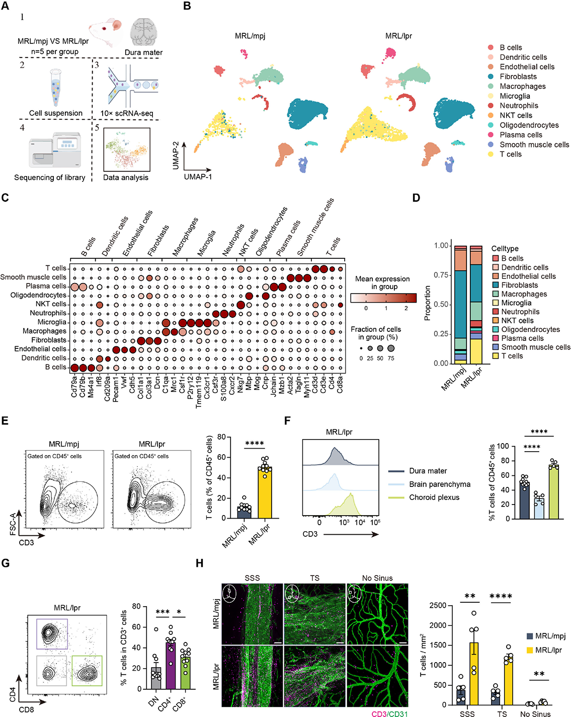
Single-cell transcriptomic profiling reveals a pro-inflammatory immune landscape characterized by T cell accumulation in the dura mater of MRL/lpr mice. A Schematic workflow of the experimental design for single-cell RNA sequencing (scRNA-seq) of dural cells in 16 weeks old MRL/lpr and MRL/mpj mice. B Uniform Manifold Approximation and Projection (UMAP) visualization of the major cell types identified in the dura mater, split by group. C Dot plot showing the expression of canonical marker genes used to identify cell clusters. D Stacked bar plot illustrating the relative proportions of immune cell subtypes in MRL/mpj and MRL/lpr mice. E Representative flow cytometry contour plots and quantification of T cells (CD3+) as a percentage of total CD45+ cells in the dura mater. F Representative histograms and quantitative comparison of T cell proportions (within CD45+ cells) across three CNS compartments: dura mater, brain parenchyma, and choroid plexus in MRL/lpr mice. G Representative flow cytometry plots and quantification of T cell subsets (CD4+, CD8+, and Double-Negative [DN]) within the dural CD3+ population in MRL/lpr mice. H Representative whole-mount immunofluorescence images and quantification of T cell density in the superior sagittal sinus (SSS), transverse sinus (TS), and non-sinus regions. Scale bars: 100 μm. Data are presented as mean ± SEM. * P < 0.05, ** P < 0.01, *** P < 0.001 and **** P < 0.0001; ns, not significant by Student’s t -test in (E, H) and one-way ANOVA (F, G).
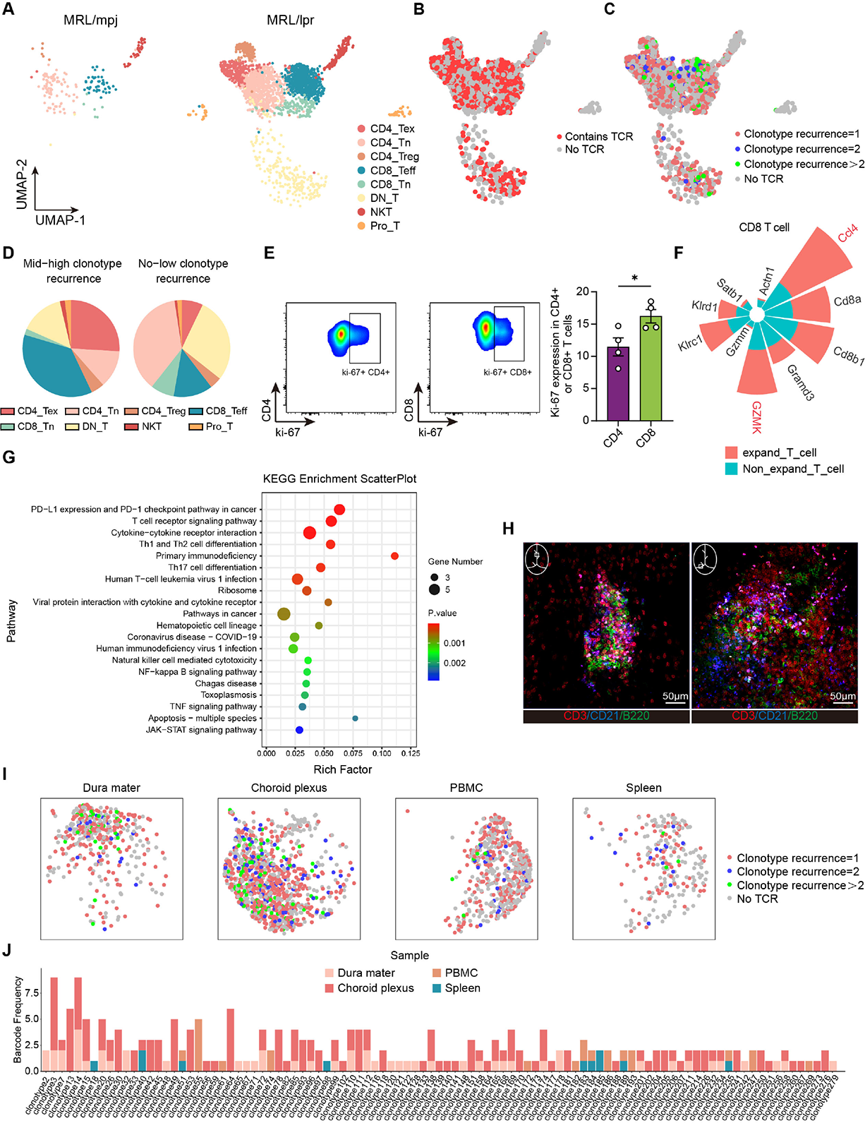
Clonal expansion of CD8+ T cells in dura mater of MRL/lpr mice. A UMAP visualization of T cell subclusters in the dura mater of MRL/mpj and MRL/lpr mice. B UMAP projection showing the recovery of T cell receptor (TCR) sequences. C Clonal space analysis on the UMAP projection. Cells are colored by clonotype recurrence. D Pie charts illustrating the composition of T cell subsets within the highly expanded (Mid-high clonotype recurrence) versus non-expanded (No-low clonotype recurrence) repertoires. E Representative flow cytometry contour plots and quantification of Ki-67 expression in dural CD4+ and CD8+ T cells. F Polar bar plot displaying the top differentially expressed genes between clonally expanded and non-expanded T cells. G KEGG pathway enrichment analysis of upregulated genes in the clonally expanded T cell population. H Representative whole-mount immunofluorescence images of dural tertiary lymphoid structures (TLS). Staining for T cells (CD3, red), follicular dendritic cells (CD21, blue), and B cells (B220, green). Scale bars: 100 μm. I UMAP plots split by tissue origin (Dura mater, Choroid plexus, PBMC, and Spleen), colored by clonotype recurrence. J Barcode frequency plot tracking the distribution of specific clonotypes across different tissues. The dura mater shares abundant clonotypes with the choroid plexus. Data are presented as mean ± SEM. * P < 0.05, ** P < 0.01, *** P < 0.001 and **** P < 0.0001; ns, not significant by Student’s t -test in (E).
REFERENCES: NIL.
Acknowledgments: NIL.
Disclosure of Interests: None declared.