fetching data ... 
Background: Systemic lupus erythematosus (SLE) is driven by pathogenic B cell activation and autoantibody production. Oxidative stress and lipid metabolic abnormalities have been implicated in SLE [1], yet clinically relevant lipid pathways connecting oxidative stress to B cell hyperactivity remain insufficiently defined.
Objectives: To characterize oxidative stress–associated phospholipid/lysophospholipid alterations in SLE and evaluate their associations with disease activity and B cell activation, with preliminary therapeutic validation via PLA2 inhibition [2].
Methods: Peripheral blood lipidomics was performed in SLE (n=10) and healthy controls (HC, n=10) using non-targeted mass spectrometry, followed by targeted quantification of key lysophospholipids. PLA2, oxidized LDL (ox-LDL) and PBMC reactive oxygen species (ROS) were assessed in an independent cohort (SLE n=27, HC n=15). Associations with SLEDAI, anti-dsDNA, and complement C3 were analyzed. In vitro, purified B cells were stimulated with LPC 18:0 to assess plasma cell differentiation. In vivo, lupus-prone MRL/lpr mice received the PLA2 inhibitor darapladib or vehicle for 4 weeks, and serological, immunophenotypic, and renal outcomes were evaluated.
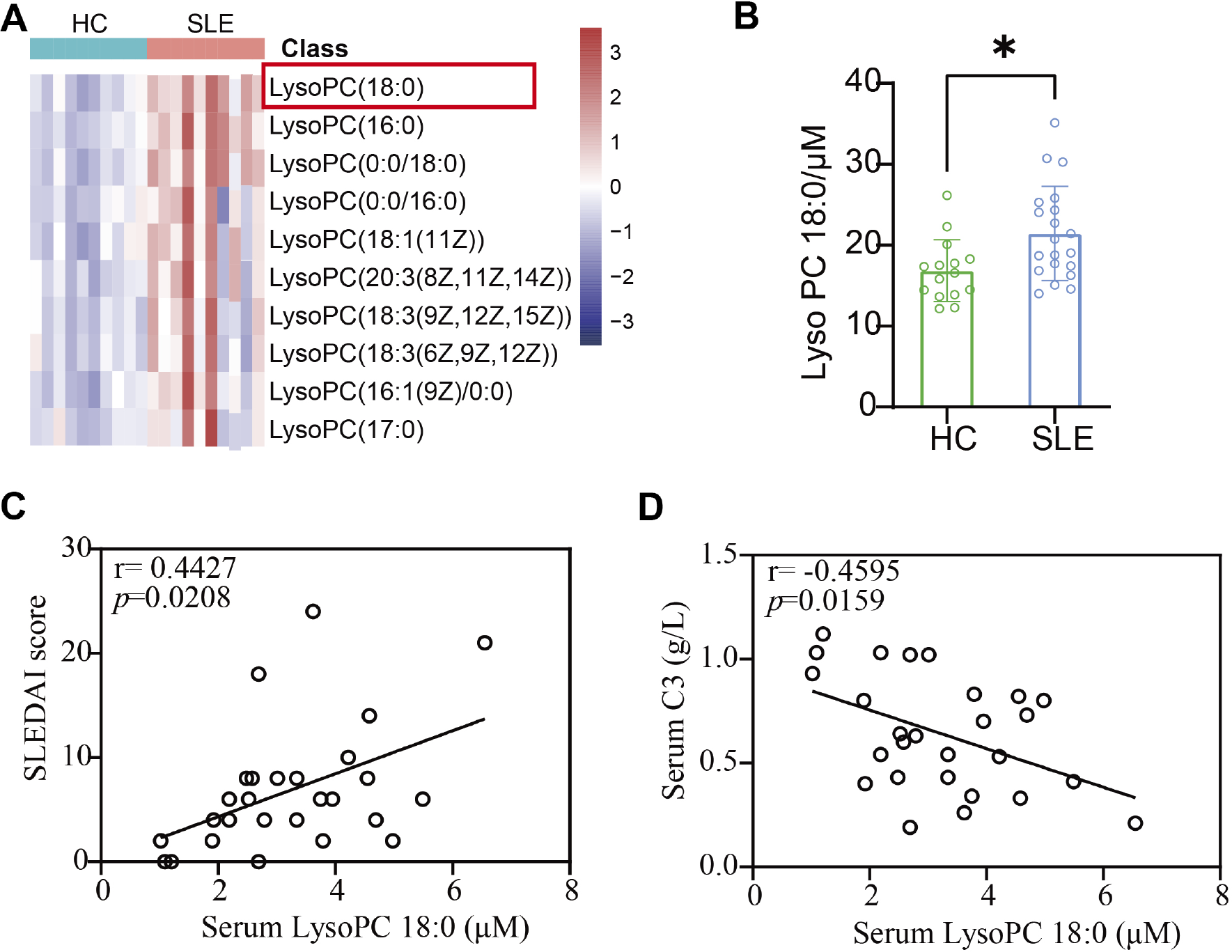
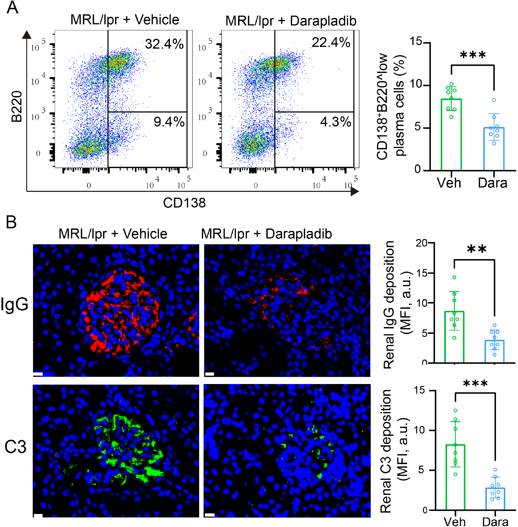
Results: Non-targeted lipidomics identified broad upregulation of lysophospholipids in SLE, with LPC 18:0 as the most abundant and discriminatory species. Targeted quantification confirmed elevated LPC 18:0 in SLE versus HC (24.4 vs 16.8 μM, p=0.012), which correlated with higher SLEDAI (r=0.442, p=0.021) and lower C3 (r=−0.456, p=0.016). Circulating PLA2 was increased in SLE (3.0 vs 1.3 μg/mL, p<0.001) and correlated with SLEDAI (r=0.567, p=0.004) and anti-dsDNA (r=0.526, p=0.008). Oxidative stress markers were also elevated (ox-LDL 394 vs 156 ng/mL, p<0.001; PBMC ROS 9.54% vs 0.89%, p<0.001). Functionally, LPC 18:0 increased plasma cell differentiation in vitro (15.3% vs 9.2%, p=0.016). In MRL/lpr mice, darapladib reduced lymphoid organ enlargement, decreased plasma cells and autoantibody levels (anti-dsDNA 337 vs 910 IU/mL, p=0.014), and alleviated renal inflammation and immune complex deposition.
Conclusions: SLE is characterized by an oxidative stress–associated PLA2–lysophospholipid signature, prominently involving LPC 18:0, which tracks with disease activity and supports B cell pathogenic differentiation. Pharmacological inhibition of PLA2 mitigates lupus manifestations in vivo, supporting PLA2-driven lipid dysregulation as a clinically relevant and therapeutically actionable pathway in SLE.
Identification of LysoPC 18:0 as a clinically relevant lysophospholipid in treatment-naïve systemic lupus erythematosus.
(A) Heatmap of the top 10 differentially abundant lysophospholipids identified by non-targeted lipidomics (SLE n=10; HC n=10), with LysoPC 18:0 highlighted.
(B) Targeted quantification of LysoPC 18:0 in SLE (n=27) and healthy controls (HC, n=15).
(C) Correlation between LysoPC 18:0 levels and SLE disease activity (SLEDAI; Spearman correlation).
(D) Correlation between ysoLPC 18:0 levels and serum complement C3 levels (Spearman correlation).
Proof-of-concept efficacy of PLA2 inhibition in a lupus-prone mouse model.
MRL/lpr mice were treated with the PLA2 inhibitor darapladib or vehicle for 4 weeks.
(A) Splenic antibody-secreting cells (ASCs) assessed by flow cytometry using B220 and CD138. Representative B220 × CD138 plots from vehicle- and darapladib-treated mice are shown, with quantification of CD138 + B220^low plasma cells.
(B) Renal immune complex deposition evaluated by immunofluorescence staining of IgG and complement C3. Representative images from vehicle- and darapladib-treated mice are shown, with quantification of renal IgG/C3 deposition expressed as mean fluorescence intensity (MFI, a.u.).
In addition, peripheral anti-dsDNA antibody titers were measured by ELISA and were reduced in darapladib-treated mice. Data are presented as mean ± SEM. Statistical significance was assessed using unpaired t-tests.
REFERENCES: [1] Perl A. Oxidative stress in the pathology and treatment of systemic lupus erythematosus. Nat Rev Rheumatol . 2013;9(11):674-686.
[2] Lu L, Hu C, Zhao Y, He L, Zhou J, Li H, et al. Shotgun lipidomics revealed altered profiles of serum lipids in systemic lupus erythematosus closely associated with disease activity. Biomolecules . 2018;8(4):105.
Acknowledgments: NIL.
Disclosure of Interests: None declared.